

Introduction
Brain health is an integral aspect of overall well-being, yet many conditions affecting the brain remain poorly understood by the general public. Among these, brain scarring is a complex and often concerning phenomenon. The presence of MRI spots on the brain, indicative of scarred brain tissue, can be alarming to both patients and medical professionals. While some brain scarring may be benign, others may signal underlying neurological conditions that require careful monitoring and intervention.
You may also like : Best Things for Brain Health: Expert-Backed Strategies to Keep Your Mind Sharp
This article explores the causes of brain scarring, its implications for cognitive function and long-term health, and the role of medical imaging in detecting spots on the brain. By examining the available scientific literature and expert opinions, we aim to provide a comprehensive understanding of how brain scarring occurs, how it is diagnosed, and what can be done to mitigate its long-term effects. This information is crucial for those seeking to optimize brain health and longevity while navigating concerns related to neurological well-being.
What is Brain Scarring?
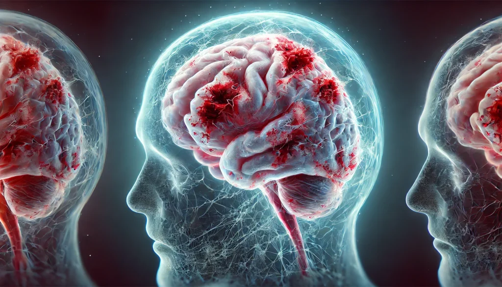
Brain scarring, also known as gliosis, occurs when the brain responds to injury or disease by forming scar tissue. Unlike scars on the skin, which serve as protective barriers, scar tissue in the brain can disrupt neural communication and lead to functional impairments. The brain’s unique structure and function mean that even small areas of scarring can have profound effects on cognition, movement, and overall neurological health.
The brain’s response to injury is mediated by specialized glial cells, primarily astrocytes, which proliferate in response to damage. This reaction, called reactive gliosis, leads to the formation of scar-like tissue that can interfere with normal neural function. In many cases, this scarring is detected as MRI spots on the brain, which may appear as bright or dark areas depending on the imaging modality used. While some instances of brain scarring are asymptomatic, others may be linked to progressive neurological diseases, making early detection and proper interpretation of MRI findings essential.
Causes of Brain Scarring
Brain scarring can arise from a variety of causes, ranging from acute trauma to chronic neurological conditions. Understanding these causes is key to both prevention and management of scarred brain tissue.
Traumatic Brain Injury (TBI)
One of the most common causes of brain scarring is traumatic brain injury (TBI), which can result from accidents, falls, sports injuries, or violent impacts. When the brain experiences a sudden jolt or blow, neurons and glial cells may be damaged, triggering an inflammatory response. This inflammation, while a natural part of the healing process, can lead to the formation of scarred brain tissue that persists long after the initial injury has healed. Repeated head trauma, such as that seen in contact sports, can exacerbate scarring and contribute to neurodegenerative conditions like chronic traumatic encephalopathy (CTE).
Stroke and Vascular Damage
Stroke, whether ischemic (caused by a blood clot) or hemorrhagic (caused by bleeding in the brain), often results in localized brain damage and subsequent scarring. When blood flow to the brain is interrupted, neurons in the affected area die, leading to the formation of MRI spots on the brain. Over time, this damaged tissue may be replaced by glial scarring, which can impair recovery and contribute to post-stroke cognitive and motor deficits.
Multiple Sclerosis and Autoimmune Disorders
Multiple sclerosis (MS) is a chronic autoimmune disease that leads to the destruction of myelin, the protective covering of nerve fibers. The resulting damage is visible on MRI scans as spots on the brain, which correspond to areas of demyelination and scarring. In MS, the immune system mistakenly attacks the central nervous system, leading to recurring episodes of inflammation and neurodegeneration. Other autoimmune conditions, such as lupus and neuromyelitis optica, can also cause brain scarring by triggering inflammatory processes that damage neural tissue.
Neuroinfections
Certain infections, including bacterial meningitis, viral encephalitis, and Lyme disease, can cause inflammation in the brain, leading to scarring. Infections of the central nervous system often provoke a strong immune response, which, while necessary for fighting pathogens, can also result in collateral damage to healthy brain tissue. This damage may manifest as MRI spots on the brain, signaling areas of past or ongoing inflammation.
Neurodegenerative Diseases
Conditions such as Alzheimer’s disease, Parkinson’s disease, and amyotrophic lateral sclerosis (ALS) are associated with progressive neural damage and scarring. As neurons degenerate, the brain attempts to compensate by forming scar tissue, which can contribute to further functional decline. In Alzheimer’s disease, for example, the accumulation of amyloid plaques and tau tangles leads to widespread neuronal loss, accompanied by reactive gliosis and scarring in affected regions.
Chronic Inflammation and Aging
Aging is a natural process that can contribute to brain scarring, particularly when chronic inflammation is present. Factors such as poor diet, lack of exercise, and exposure to environmental toxins can exacerbate neuroinflammation, increasing the risk of developing spots on the brain as detected by MRI. Additionally, conditions such as diabetes and hypertension, which are more prevalent in older adults, can lead to vascular damage that promotes brain scarring over time.
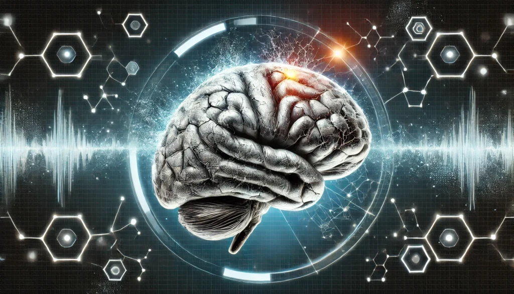
Diagnosing Brain Scarring with MRI
MRI (magnetic resonance imaging) is a crucial tool for detecting scarred brain tissue and assessing its impact on neurological function. Different types of MRI scans, including T1-weighted, T2-weighted, and FLAIR (fluid-attenuated inversion recovery), can reveal distinct features of brain scarring.
Understanding MRI Spots on the Brain
When examining MRI spots on the brain, radiologists look for patterns that distinguish benign findings from those indicative of disease. For instance, white matter hyperintensities (WMHs) are common in aging individuals and may not always be clinically significant. However, clusters of spots, especially in younger patients, may raise concerns about conditions like multiple sclerosis or small vessel disease.
Differentiating Between Normal Variants and Pathological Findings
Not all spots on the brain seen on MRI scans indicate a serious condition. Some findings may be incidental and unrelated to any underlying disease. However, when scarring appears in conjunction with symptoms such as cognitive decline, motor dysfunction, or recurrent headaches, further evaluation is warranted.
Long-Term Effects of Brain Scarring
Brain scarring can have a range of long-term effects depending on its severity and location. Some individuals may experience no noticeable symptoms, while others may develop cognitive, motor, or psychological impairments.
Cognitive Decline and Memory Impairment
Scarring in areas associated with memory, such as the hippocampus, can contribute to conditions like dementia and mild cognitive impairment (MCI). Even in cases where scarring is minimal, disrupted neural connectivity can lead to subtle deficits in attention, processing speed, and problem-solving abilities.
Motor and Sensory Deficits
Scarring in the motor cortex or cerebellum can result in movement disorders, balance issues, and coordination problems. Similarly, scarring in sensory-processing regions may lead to altered perception of touch, pain, or temperature.
Psychological and Emotional Effects
Depression, anxiety, and mood instability have been linked to brain scarring, particularly when it affects regions involved in emotional regulation. Damage to the prefrontal cortex, for instance, can impair decision-making and impulse control, contributing to behavioral changes.
Frequently Asked Questions (FAQ) About Brain Scarring
1. Can brain scarring heal over time, or is it permanent?
Brain scarring, also known as gliosis, is generally considered permanent because the nervous system lacks the regenerative capacity of other tissues like skin or muscle. Unlike other forms of healing, where damaged tissue is replaced with functional cells, scarred brain tissue disrupts neural connectivity rather than restoring it. However, some level of neuroplasticity allows the brain to compensate by forming new connections around the damaged area. While certain treatments, including cognitive rehabilitation and neurostimulation, may improve function, the underlying brain scarring does not typically disappear. Research into regenerative medicine, including stem cell therapy and neurotrophic factors, is ongoing and offers hope for future treatments.
2. What are the most common symptoms associated with brain scarring?
The symptoms of brain scarring vary depending on the affected region. If scarring occurs in the frontal lobe, individuals may experience difficulties with decision-making, impulse control, and social behavior. When scarred brain tissue affects motor regions, movement disorders, muscle weakness, or coordination problems may arise. MRI spots on the brain often correlate with cognitive issues, such as memory loss, difficulty concentrating, or slowed information processing. In some cases, people with spots on the brain may develop mood disorders, including depression and anxiety, due to altered neurotransmitter activity. While symptoms may progress gradually, early intervention and cognitive therapies can help mitigate the impact.
3. Do MRI spots on the brain always indicate serious neurological conditions? Not necessarily. MRI spots on the brain can have a range of causes, from benign age-related changes to serious neurological diseases like multiple sclerosis (MS) or small vessel disease. Some findings are incidental and pose no immediate health risk, while others require closer monitoring to assess their progression. The context in which spots on the brain appear—such as whether they are accompanied by symptoms—plays a crucial role in determining their significance. Physicians rely on additional tests, including blood work, spinal taps, and clinical assessments, to differentiate between harmless anomalies and more concerning conditions. Regular follow-ups and lifestyle changes, such as maintaining cardiovascular health, can help prevent further deterioration.
4. How does aging contribute to brain scarring?
Aging is a natural risk factor for brain scarring, largely due to the cumulative effects of vascular damage, inflammation, and reduced neurogenesis. Over time, tiny blood vessels in the brain may become less efficient, leading to minor strokes or ischemic damage that results in scarred brain tissue. Additionally, chronic low-level inflammation, often linked to conditions such as diabetes and hypertension, exacerbates cellular damage and promotes scarring. Many older adults develop MRI spots on the brain as a result of these processes, although not all experience significant cognitive decline. Preventative measures, including a brain-healthy diet rich in antioxidants, regular exercise, and cognitive engagement, can help slow age-related neurological changes.
5. Can lifestyle choices reduce the risk of developing brain scarring?
Yes, lifestyle choices play a critical role in reducing the likelihood of brain scarring and its associated complications. Maintaining cardiovascular health is particularly important, as high blood pressure, high cholesterol, and diabetes are linked to vascular damage that contributes to scarred brain tissue. Regular physical activity improves blood flow to the brain and supports neuroplasticity, helping to counteract the effects of aging and injury. A diet rich in omega-3 fatty acids, antioxidants, and anti-inflammatory foods can help protect against neuroinflammation that leads to spots on the brain. Additionally, mental stimulation through learning, problem-solving, and social engagement fosters resilience in neural networks, potentially reducing the impact of existing scarring.
6. Is there a link between brain scarring and neurodegenerative diseases? Yes, brain scarring is commonly associated with neurodegenerative diseases such as Alzheimer’s, Parkinson’s, and amyotrophic lateral sclerosis (ALS). In these conditions, ongoing neuronal loss and inflammation lead to the formation of scarred brain tissue, which further impairs cognitive and motor functions. MRI spots on the brain seen in neurodegenerative disorders often represent areas of previous injury or chronic damage caused by toxic protein buildup, vascular insufficiency, or immune system dysfunction. While scarring itself may not directly cause disease, it can be a marker of underlying pathology that contributes to progressive decline. Early detection and intervention, including lifestyle changes and medication, can help slow disease progression and improve quality of life.
7. How does traumatic brain injury (TBI) contribute to brain scarring? Traumatic brain injury (TBI) is one of the leading causes of brain scarring, often resulting from falls, sports injuries, or accidents. When the brain experiences a forceful impact, neurons and glial cells are damaged, triggering an inflammatory response. In some cases, scarred brain tissue forms in response to this injury, leading to long-term cognitive, motor, and emotional difficulties. People with a history of multiple concussions, such as athletes in contact sports, are at increased risk of developing chronic traumatic encephalopathy (CTE), a condition characterized by widespread MRI spots on the brain. Treatment focuses on symptom management, neurorehabilitation, and strategies to reduce further injury, such as protective headgear and avoiding high-risk activities.
8. Can brain scarring be reversed with medication or therapy?
At present, there are no medications that can completely reverse brain scarring, but certain treatments can help manage its effects. Anti-inflammatory drugs, neuroprotective agents, and medications that improve blood flow to the brain may reduce further damage and support recovery. Cognitive therapy and rehabilitation techniques can help individuals adapt by strengthening unaffected neural pathways. In some experimental studies, stem cell therapy and brain stimulation techniques have shown promise in promoting functional recovery in areas affected by scarred brain tissue. However, these treatments are still in early stages, and more research is needed to determine their long-term effectiveness.
9. Are there specific medical conditions that increase the likelihood of MRI spots on the brain?
Yes, several medical conditions increase the likelihood of MRI spots on the brain. Multiple sclerosis, small vessel disease, lupus, and certain types of migraines are frequently associated with spots on the brain seen on MRI scans. Additionally, chronic hypertension, diabetes, and autoimmune disorders that trigger systemic inflammation can contribute to scarred brain tissue. Infections such as meningitis or encephalitis may also leave behind visible scarring detectable through neuroimaging. Because these conditions affect brain health in different ways, treatment strategies vary, and early medical intervention is often crucial to managing long-term outcomes.
10. What are the latest advancements in research on brain scarring? Advancements in neuroscience and medical technology are providing new insights into brain scarring and its potential treatments. Emerging research in regenerative medicine is exploring how stem cells can be used to replace damaged neural tissue and improve brain function. Gene therapy is another promising avenue, with scientists investigating ways to regulate the inflammatory responses that contribute to scarred brain tissue. Non-invasive brain stimulation techniques, such as transcranial magnetic stimulation (TMS) and direct current stimulation, are being tested for their ability to enhance neuroplasticity and repair damaged connections. Additionally, advanced imaging techniques are improving the ability to detect and monitor MRI spots on the brain, allowing for earlier diagnosis and more personalized treatment approaches. While these therapies are still in development, they offer hope for future breakthroughs in managing and potentially reversing the effects of brain scarring.
Conclusion
Brain scarring is a complex phenomenon with diverse causes and consequences. Whether resulting from injury, disease, or aging, scarred brain tissue can have profound implications for cognitive and neurological health. Understanding the causes of MRI spots on the brain and their potential impact is essential for early intervention and management. By prioritizing brain health through lifestyle choices, medical monitoring, and proactive care, individuals can reduce the risk of long-term neurological decline and enhance overall well-being.
neurological health, brain lesion causes, cognitive impairment risk, neuroplasticity and healing, white matter changes, brain MRI interpretation, neurological inflammation, stroke recovery insights, head trauma effects, multiple sclerosis symptoms, aging and brain function, vascular health and cognition, autoimmune brain disorders, encephalitis aftereffects, chronic headaches and brain health, neurodegeneration prevention, traumatic brain injuries, dementia early signs, nervous system repair, brain rehabilitation strategies
Further Reading:
What side effects can radiation therapy on the brain cause?
The chronic and evolving neurological consequences of traumatic brain injury
MRI Brain for Traumatic Brain Injury
Disclaimer
The information contained in this article is provided for general informational purposes only and is not intended to serve as medical, legal, or professional advice. While Health11News strives to present accurate, up-to-date, and reliable content, no warranty or guarantee, expressed or implied, is made regarding the completeness, accuracy, or adequacy of the information provided. Readers are strongly advised to seek the guidance of a qualified healthcare provider or other relevant professionals before acting on any information contained in this article. Health11News, its authors, editors, and contributors expressly disclaim any liability for any damages, losses, or consequences arising directly or indirectly from the use, interpretation, or reliance on any information presented herein. The views and opinions expressed in this article are those of the author(s) and do not necessarily reflect the official policies or positions of Health11News.
