
")
Introduction: The Marvel of the Human Brain
The human brain is an extraordinary organ, responsible for processing all of the information your brain receives and enabling every function essential to life. As the control center of the body, it is often described as the most complex biological structure known to science. Understanding the different parts of the brain, including its anatomy and specific lobes, provides crucial insight into cognitive functions, memory, sensory processing, and movement. A detailed brain diagram or brain labeled diagram reveals the intricate organization of this vital organ, helping to highlight the relationships between different regions and their specialized functions.
You may also like : Best Things for Brain Health: Expert-Backed Strategies to Keep Your Mind Sharp
With advanced neuroimaging techniques, scientists have gained a clearer understanding of where in the brain most information processing occurs and how various brain structures interact. This knowledge is fundamental for medical professionals, neuroscientists, and anyone interested in brain health and cognitive enhancement. In this article, we will explore the anatomy of the brain, including the lobes of the brain, what part of the brain controls movement, what part of the brain helps with organization, and much more. We will also examine whether there is muscle around your brain and answer the common question: Is the brain a muscle or organ?
By examining each section in depth, we will gain a comprehensive understanding of what part of the brain controls simple movements, what part of the brain helps you perceive things, and how different lobes contribute to various functions such as speech, taste, memory, and sight. Whether you are a student, a professional, or simply curious about the inner workings of your mind, this guide will provide valuable insights into the fascinating world inside of a brain.
The Structure of the Brain: An Overview
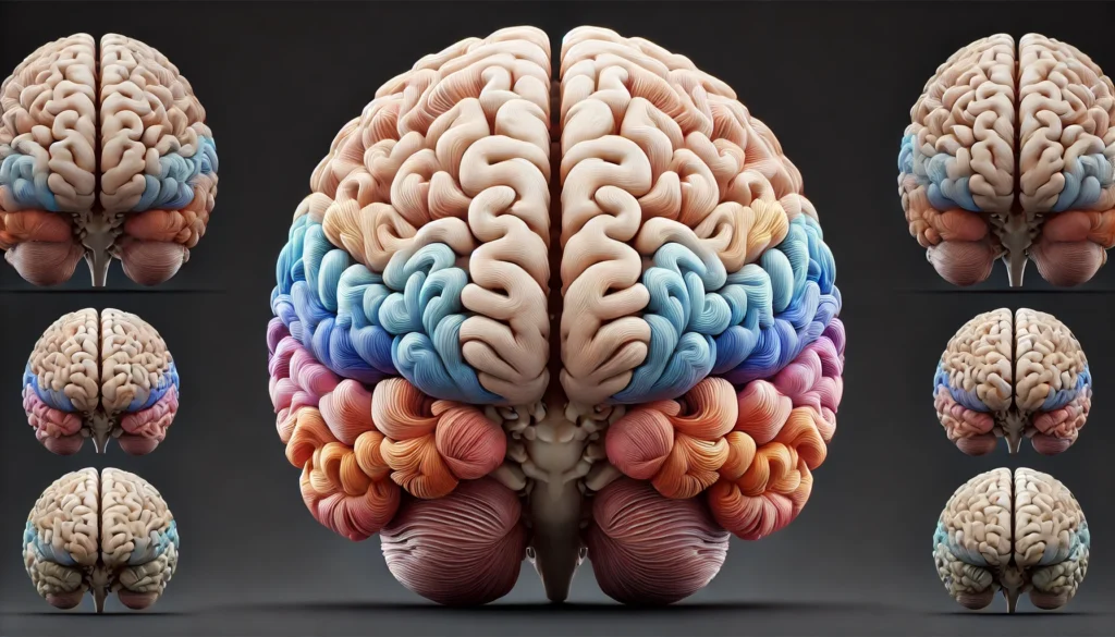
The brain is divided into several major regions, each with distinct roles in cognition, motor control, and sensory processing. At a high level, the brain is composed of the cerebrum, cerebellum, and brainstem. The cerebrum is the biggest part of the brain and is responsible for higher cognitive functions such as reasoning, decision-making, and voluntary movement. The cerebellum, often referred to as “this brain structure that deals with balance and muscle memory,” is essential for coordinating movement and maintaining equilibrium. The brainstem connects the brain to the spinal cord and controls vital functions such as breathing and heart rate.
The cerebrum is further divided into two hemispheres, which are connected by the corpus callosum, a thick bundle of nerve fibers that facilitates communication between the left and right sides of the brain. Each hemisphere is responsible for controlling specific functions, often in a lateralized manner. For instance, what side of the brain controls speech? The left hemisphere is primarily responsible for language processing and speech production, whereas the right hemisphere is more involved in spatial reasoning and creativity. Understanding what part of the brain controls eyesight, taste, and other sensory experiences helps to elucidate the intricate mechanisms that underpin human perception.
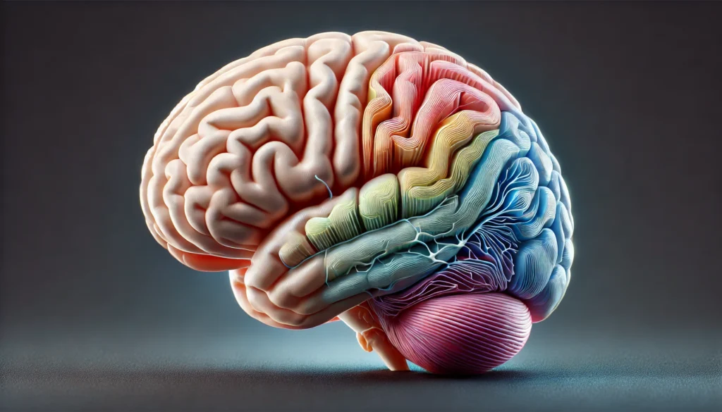
A side view of the brain labeled in an anatomical picture of the brain reveals the major lobes that contribute to various cognitive and sensory functions. Each lobe has specialized areas that work together to form a coherent and efficient system, processing external stimuli and generating appropriate responses. These parts of the brain are essential for daily activities, from recognizing faces to planning future actions.
The Lobes of the Brain and Their Functions
The cerebrum is divided into four primary lobes, each with distinct roles in cognitive function, sensory processing, and voluntary movement. These lobes are the frontal lobe, parietal lobe, temporal lobe, and occipital lobe. Each lobe houses specific regions responsible for different aspects of perception, memory, and control.
The Frontal Lobe: The Center of Decision-Making and Movement
The frontal lobe is the largest and most advanced part of the brain, located at the front of the cerebrum. It plays a critical role in executive functions such as reasoning, problem-solving, decision-making, and emotional regulation. This region also contains the primary motor cortex, which controls voluntary movements and answers the question: what part of brain controls movement?
Another essential function of the frontal lobe is organization. Many ask, “What part of the brain helps with organization?” The answer lies within the prefrontal cortex, a subregion of the frontal lobe responsible for planning, organizing thoughts, and maintaining focus. Additionally, the Broca’s area, located in the left frontal lobe, is essential for speech production, reinforcing the role of the left hemisphere in language processing.
The Parietal Lobe: Processing Sensory Information
Positioned behind the frontal lobe, the parietal lobe processes sensory input related to touch, temperature, and pain. It is responsible for spatial awareness and navigation, making it crucial for interpreting sensory data and understanding the surrounding environment.
One key question often asked is: “What part of the brain helps you perceive things?” The answer lies in the parietal lobe, particularly in the somatosensory cortex. This region interprets tactile sensations and integrates information from different sensory modalities, allowing individuals to recognize objects, textures, and spatial relationships.
The Temporal Lobe: Memory and Auditory Processing
The temporal lobe, located on the sides of the brain, plays a crucial role in auditory processing, language comprehension, and memory formation. This region contains the hippocampus, which is central to memory consolidation. A common query regarding brain function is: “What part of the cerebrum controls memory?” The hippocampus and surrounding medial temporal structures are essential for converting short-term memories into long-term ones.
Furthermore, the temporal lobe contains Wernicke’s area, which is responsible for language comprehension. Damage to this area can result in difficulties understanding spoken and written language, highlighting its importance in communication.
The Occipital Lobe: Vision and Perception
The occipital lobe, located at the back of the brain, is primarily responsible for processing visual information. It answers the questions “What part of the brain controls sight?” and “What part of the brain controls eyesight?” by housing the primary visual cortex, where signals from the retina are interpreted. Damage to this area can lead to visual impairments, such as difficulty recognizing faces or interpreting spatial relationships.
Frequently Asked Questions About Brain Anatomy and Function
1. What is the best word for how your brain works?
The best word for how your brain works depends on the context in which you are describing its functions. In scientific terms, “cognition” refers to the mental processes involved in acquiring knowledge and understanding. If you are focusing on how the brain processes sensory input and coordinates responses, “neurofunction” might be appropriate. When discussing the brain’s ability to adapt and reorganize itself, the term “neuroplasticity” is widely used. The way the brain integrates all of the information your brain receives is often referred to as “cognitive processing.” Ultimately, the complexity of the brain’s operations means that multiple terms may be necessary to fully capture how it works.
2. Are there different ways to view the brain in medical imaging?
Yes, there are several ways to view the brain in medical imaging, each providing different insights into brain anatomy and function. MRI (Magnetic Resonance Imaging) offers highly detailed images, often used to visualize the lobes of the brain and deeper structures such as the hippocampus. A CT (Computed Tomography) scan provides cross-sectional images, useful for detecting brain injuries. Functional MRI (fMRI) highlights areas where most information processing occurs by tracking blood flow changes. PET (Positron Emission Tomography) scans detect metabolic activity, helping to identify issues such as dementia. An anatomical picture of the brain obtained through these methods can provide a comprehensive view of brain health and function.
3. Is the brain a muscle or organ?
The brain is an organ, not a muscle, despite its ability to “work out” through mental exercises. Unlike muscles, which contract to produce movement, the brain is composed of neurons and glial cells that transmit electrical and chemical signals. While there is no muscle around your brain, the brain is encased in a protective layer of connective tissue called the meninges. Brain function can be improved through cognitive training, much like muscles grow stronger through exercise. Maintaining brain health through mental stimulation, physical activity, and proper nutrition is essential for long-term cognitive performance.
4. What part of the brain controls taste?
Taste perception is controlled by the gustatory cortex, which is located in the insular and frontal operculum regions of the brain. When you eat food, sensory receptors on your tongue send signals to the brainstem, which then relays information to the thalamus. The thalamus processes this information and directs it to the gustatory cortex, allowing you to perceive flavors. Additionally, the limbic system plays a role in associating taste with emotions and memories. Understanding what part of the brain controls taste helps in studying disorders that affect flavor perception, such as anosmia and ageusia.
5. Where in the brain does most information processing occur?
Most information processing occurs in the cerebral cortex, the outermost layer of the brain. This highly folded structure contains specialized areas responsible for different cognitive functions, such as the prefrontal cortex for decision-making and the occipital lobe for visual processing. The parietal lobe integrates sensory data to help you understand spatial relationships and body positioning. The temporal lobe is essential for memory formation and auditory processing, while the frontal lobe plays a significant role in problem-solving and executive functions. A brain labeled diagram highlights these regions, providing a clearer understanding of their roles in cognition.
6. What part of the brain helps with organization and planning?
The prefrontal cortex, located in the frontal lobe, is responsible for organization, planning, and decision-making. It allows individuals to set goals, prioritize tasks, and regulate impulses, making it essential for executive function. Damage to this area can lead to difficulties in maintaining focus, managing time, and controlling behavior. The dorsolateral prefrontal cortex is particularly crucial in working memory, which helps in keeping track of multiple pieces of information at once. Research into this region has provided insights into disorders such as ADHD, where organizational skills are often impaired.
7. What is the biggest part of the brain, and what does it do?
The biggest part of the brain is the cerebrum, which makes up about 85% of the brain’s weight. It is divided into two hemispheres, each controlling opposite sides of the body. The cerebrum is responsible for higher cognitive functions, including thought, language, memory, and voluntary movement. The different lobes of the brain within the cerebrum specialize in various tasks, such as the occipital lobe for vision and the temporal lobe for auditory processing. Studying brain anatomy labeled with different functional areas helps scientists understand how the cerebrum governs complex behaviors and emotions.
8. What part of the cerebrum controls memory?
Memory is primarily controlled by the hippocampus, a structure located in the medial temporal lobe of the cerebrum. The hippocampus is crucial for converting short-term memories into long-term ones, allowing for the retention of new information. Damage to this area, as seen in conditions like Alzheimer’s disease, can severely impact memory formation. Other regions, such as the prefrontal cortex, play a role in working memory and decision-making. Research into the hippocampus and its role in memory has been instrumental in understanding neurodegenerative diseases and potential treatments.
9. What part of the brain controls simple movements?
Simple movements are controlled by the primary motor cortex, which is located in the frontal lobe. This area sends signals to muscles through the spinal cord, enabling voluntary movement. The basal ganglia, a group of structures deep inside the brain, help regulate movement and prevent involuntary muscle contractions. The cerebellum, often described as “this brain structure deals with balance and muscle memory,” refines motor actions to ensure smooth coordination. Disorders affecting these regions, such as Parkinson’s disease, can lead to tremors and difficulties with movement control. Studying a brain diagram can help visualize the pathways involved in motor function.
10. What side of the brain controls speech?
In most people, speech is controlled by the left hemisphere of the brain, specifically in areas known as Broca’s area and Wernicke’s area. Broca’s area, located in the frontal lobe, is responsible for speech production, allowing individuals to form words and articulate sentences. Wernicke’s area, found in the temporal lobe, is essential for language comprehension, ensuring that speech makes sense. Damage to these areas can result in language impairments such as aphasia, where speech production or comprehension is affected. A side view of the brain labeled with these regions helps illustrate how they contribute to communication and language processing.
Conclusion: The Brain’s Complexity and Functionality
Understanding the anatomy of the brain is essential for comprehending how cognitive functions, sensory processing, and motor control work together to create human experience. Each lobe of the brain contributes uniquely to perception, movement, and memory, demonstrating the brain’s intricate design. By examining the various parts of the brain through an anatomical picture of the brain or a brain labeled diagram, we gain insight into how different regions interact and contribute to overall function.
The study of brain anatomy labeled with distinct functional areas offers valuable knowledge for medical professionals, researchers, and individuals interested in brain health. Whether exploring the inside of a brain to determine what part of the brain controls taste or investigating the biggest part of the brain responsible for higher cognition, ongoing research continues to uncover the complexities of this remarkable organ. Through understanding and further exploration, we can unlock the potential for cognitive enhancement, anti-aging strategies, and improved neurological health.
neuroscience basics, cognitive function explained, brain health tips, human brain structure, neural pathways and cognition, brain function overview, understanding brain processing, sensory perception in the brain, motor control in the brain, cerebral cortex functions, brain hemisphere specialization, memory and learning processes, how the brain interprets information, brain connectivity and communication, neurological health and aging, brain plasticity and adaptation, role of the prefrontal cortex, speech and language processing in the brain, visual processing and recognition, improving brain efficiency
Further Reading:
The anatomy of the brain – learned over the centuries
Understanding the Human Brain Anatomy
Neuroanatomy: Brain Structure and Function
Disclaimer
The information contained in this article is provided for general informational purposes only and is not intended to serve as medical, legal, or professional advice. While Health11News strives to present accurate, up-to-date, and reliable content, no warranty or guarantee, expressed or implied, is made regarding the completeness, accuracy, or adequacy of the information provided. Readers are strongly advised to seek the guidance of a qualified healthcare provider or other relevant professionals before acting on any information contained in this article. Health11News, its authors, editors, and contributors expressly disclaim any liability for any damages, losses, or consequences arising directly or indirectly from the use, interpretation, or reliance on any information presented herein. The views and opinions expressed in this article are those of the author(s) and do not necessarily reflect the official policies or positions of Health11News
