Understanding the Obsessive Brain: Why It Matters for Cognitive Health
Obsessive-Compulsive Disorder (OCD) is far more than the colloquial reference to excessive cleanliness or repetitive checking. It is a chronic neuropsychiatric condition marked by intrusive, distressing thoughts (obsessions) and ritualistic behaviors (compulsions) that aim to mitigate anxiety. These behaviors are not simply bad habits or personality quirks; they stem from deeply rooted neurological mechanisms. Understanding the architecture and function of the obsessive brain offers more than just insight into a specific disorder. It opens a portal to understanding how neural networks, brain chemistry, and cognitive processes interplay in both typical and atypical brain states. Recent advances in neuroimaging allow researchers to visualize these processes with unprecedented clarity, making OCD brain scan data a goldmine for neuroscience.
You may also like : Best Things for Brain Health: Expert-Backed Strategies to Keep Your Mind Sharp
The stakes are high—not only for those suffering from OCD but for the broader pursuit of cognitive longevity. As we begin to see clear distinctions in OCD brain images compared to non-OCD brains, new questions arise about the malleability of the brain, cognitive resilience, and the long-term consequences of neural overactivation. By examining the obsessive compulsive disorder brain scan results and comparing normal brain vs OCD brain structures, researchers are beginning to unravel what it means to live with an obsessive brain and how that experience might affect aging, memory, and neuroplasticity.
A Neuroscientific Overview of Obsessive-Compulsive Disorder
At its core, OCD is a disorder of cognitive regulation. The afflicted individual typically experiences intrusive thoughts that trigger excessive anxiety, prompting compulsive behaviors aimed at relieving that distress. Neuroimaging has shown that these behaviors are not random or voluntary—they correspond with specific patterns of brain activation. Brain obsessive compulsive disorder research has found significant differences in activity within the cortico-striato-thalamo-cortical (CSTC) loop, a brain circuit deeply involved in habit formation, reward anticipation, and error detection.
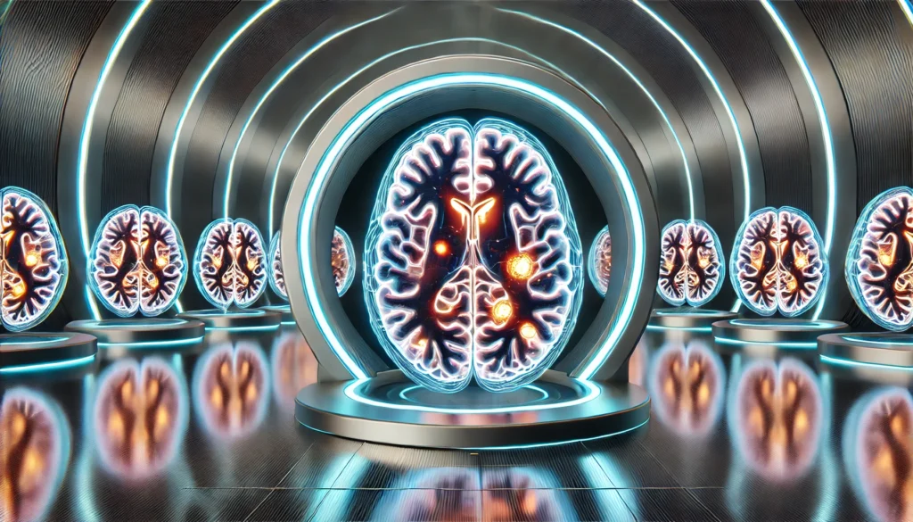
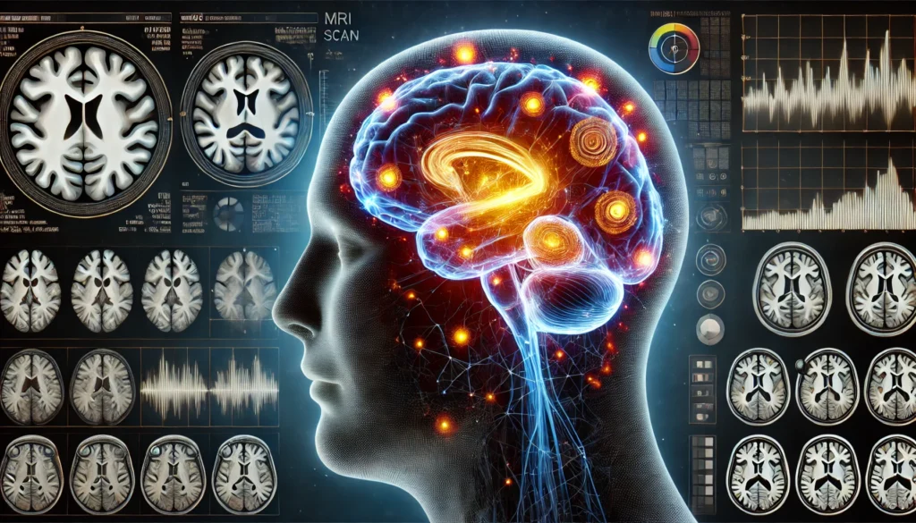
One of the most consistently implicated regions in OCD brain scans is the orbitofrontal cortex (OFC). This region, responsible for decision-making and evaluating consequences, tends to be hyperactive in those with OCD. When individuals experience obsessive thoughts, the OFC lights up with unusual intensity, indicating a maladaptive concern with perceived threats or errors. Paired with heightened activity in the anterior cingulate cortex (ACC)—which governs error detection and emotional regulation—this can create a self-reinforcing loop of anxiety and ritualistic behavior. These abnormalities are stark when comparing normal brain vs OCD brain images and suggest that the obsessive brain operates in a fundamentally different mode.
Also involved is the caudate nucleus, a structure that helps filter and prioritize incoming information. In the OCD brain, this filtering system appears impaired, resulting in a kind of neural traffic jam. Stimuli that would typically be dismissed as irrelevant continue to be processed as urgent, leading to persistent, intrusive thoughts. This lack of cognitive gating may explain why compulsions feel necessary even when the individual logically recognizes their irrationality. All these insights emphasize how obsessive compulsive disorder brain scan results help to paint a detailed picture of OCD as a systems-level dysfunction rather than a localized defect.
The Power of Imaging: What OCD Brain Scans Reveal
Modern neuroimaging techniques such as fMRI (functional Magnetic Resonance Imaging), PET (Positron Emission Tomography), and DTI (Diffusion Tensor Imaging) have revolutionized our understanding of psychiatric illnesses, including OCD. Through these tools, researchers can analyze not only brain structure but also function, connectivity, and neurotransmitter distribution. In the context of OCD, these imaging techniques reveal specific and replicable patterns of dysfunction across several neural networks.
Functional MRI studies, for example, consistently show overactivity in the OFC and ACC during tasks that simulate OCD-like decisions or when subjects are exposed to triggering stimuli. These results provide real-time visual confirmation that the obsessive brain engages differently with the environment. Furthermore, longitudinal studies using PET scans have shown that successful treatment with cognitive-behavioral therapy (CBT) or selective serotonin reuptake inhibitors (SSRIs) can normalize these patterns of overactivation. Such findings underscore the brain’s plasticity and offer hope for reversing maladaptive patterns.
Additionally, Diffusion Tensor Imaging, which maps white matter tracts in the brain, has found abnormal connectivity in OCD patients. These deviations are especially prominent in connections between the frontal cortex and deeper brain structures like the basal ganglia. The altered connectivity could explain why some individuals struggle with impulse control and emotional regulation—traits often seen in OCD. These imaging revelations not only confirm the organic basis of OCD but also inform treatment planning and early intervention strategies. Every OCD brain scan adds another piece to the puzzle, enhancing both clinical understanding and the broader framework of neuropsychiatric research.

Comparing the Normal Brain vs OCD Brain: Structural and Functional Contrasts
A compelling way to appreciate the complexities of OCD is by directly comparing a normal brain vs OCD brain through neuroimaging data. While the human brain is extraordinarily diverse, patterns begin to emerge when images from large populations are aggregated. Normal brains show balanced activity across functional networks, particularly when switching between tasks or regulating emotions. In contrast, OCD brain images reveal consistent anomalies that point to dysregulation, especially in the CSTC circuit.
In healthy individuals, the caudate nucleus effectively filters incoming stimuli, allowing the brain to move fluidly from one thought or task to another. However, in the obsessive brain, this mechanism falters, leading to excessive processing of irrelevant or distressing thoughts. This inefficiency becomes visible in OCD brain scans as persistent activation in the basal ganglia and orbitofrontal regions. The result is not merely a more active brain, but a brain that cannot properly disengage.
Another contrast lies in the Default Mode Network (DMN), a set of brain regions more active during rest or introspection. Studies show that individuals with OCD often have abnormal DMN activity, which may contribute to the relentless mental replay of obsessions. In a normal brain, the DMN deactivates during goal-directed tasks. In the OCD brain, this deactivation is often incomplete, potentially explaining the intrusive nature of obsessive thoughts. Understanding these differences is not just an academic exercise; it has real-world implications for diagnosing, treating, and potentially preventing the cognitive deterioration that can accompany chronic OCD.
Neuroplasticity and the Possibility of Change in the Obsessive Brain
One of the most hopeful revelations from OCD brain scan research is the evidence of neuroplasticity—the brain’s remarkable ability to change its structure and function in response to experience. Contrary to outdated beliefs that adult brains are relatively fixed, we now understand that targeted therapies can lead to measurable changes in brain activity and connectivity. For individuals with OCD, this means that the obsessive brain is not destiny.
CBT, particularly Exposure and Response Prevention (ERP), has been shown to alter brain function in key regions implicated in OCD. Imaging studies before and after therapy often reveal decreased activity in the OFC and ACC, correlating with symptom reduction. Similarly, pharmacological treatments like SSRIs can dampen hyperactivity in the CSTC loop, promoting greater emotional balance and impulse control. These changes are not merely symptomatic improvements—they are structural and functional modifications visible on an OCD brain scan.
Even mindfulness-based interventions have begun to show promise in reconditioning the obsessive brain. By enhancing awareness and promoting non-reactivity to intrusive thoughts, mindfulness practices may reduce DMN hyperactivity and improve frontal-limbic connectivity. These changes underscore a critical truth: the obsessive brain can heal. This plasticity not only provides hope for those with OCD but also contributes to the broader understanding of how targeted interventions can promote cognitive resilience throughout life.
Obsessive Compulsive Disorder, Aging, and Brain Longevity
The relationship between OCD and aging remains an underexplored but vitally important area of research. As individuals with OCD age, the long-term consequences of chronic neural overactivation may impact cognitive function, emotional regulation, and even physical health. Prolonged hyperactivity in regions like the OFC and ACC can contribute to neuroinflammation and oxidative stress, both of which are associated with accelerated aging.
Some longitudinal studies suggest that untreated OCD may increase the risk of developing other neuropsychiatric conditions later in life, including depression and cognitive decline. The obsessive brain, if left unchecked, may be more vulnerable to the effects of age-related neural degradation. However, proactive management of symptoms—especially through therapies that promote neuroplasticity—can potentially mitigate these risks. Effective treatment may not only reduce current distress but also serve as a buffer against future cognitive decline.
Moreover, the act of engaging in therapy itself appears to promote longevity. The structured problem-solving and emotional processing involved in CBT, for instance, can enhance executive function and support long-term brain health. Understanding the OCD brain through advanced imaging enables more precise interventions, offering a blueprint for preserving cognitive vitality even in the presence of chronic mental health challenges. In this light, the study of the obsessive compulsive disorder brain scan is not just a diagnostic tool but a roadmap to aging well.

The Broader Implications for Brain Health and Society
The insights gleaned from OCD brain scans extend beyond the individual and carry broader implications for society. As we better understand the obsessive brain, we also gain a deeper appreciation for the diverse ways in which human cognition can function. This understanding can reduce stigma and increase empathy, helping society shift from a view of OCD as a behavioral problem to one of neurobiological origin.
Furthermore, the techniques used to study the OCD brain are now being applied to other conditions, including anxiety, depression, ADHD, and even neurodegenerative diseases like Alzheimer’s. These overlapping insights may help build a more integrated understanding of mental health and brain longevity. Patterns identified in OCD brain images may illuminate vulnerabilities or compensatory mechanisms in other disorders, creating opportunities for shared treatment strategies.
There are also important policy implications. The ability to visualize mental illness through neuroimaging could strengthen the case for parity in mental and physical health care. Insurers and policymakers may be more inclined to fund evidence-based treatments when presented with concrete, visual data from obsessive compulsive disorder brain scans. These developments could revolutionize mental health care delivery, making it more personalized, predictive, and preventative.
Frequently Asked Questions: Understanding the Obsessive Brain Through Imaging
1. How do ocd brain images help differentiate OCD from other anxiety-related disorders? While traditional diagnoses rely heavily on behavioral observations and patient-reported symptoms, ocd brain images provide an objective visual framework for understanding how OCD differs neurologically from generalized anxiety disorder or panic disorder. For example, in brain obsessive compulsive disorder studies, the orbitofrontal cortex shows unique patterns of hyperactivation that aren’t commonly observed in other anxiety conditions. Additionally, increased connectivity within the cortico-striato-thalamo-cortical loop, often visible in an ocd brain scan, distinguishes OCD’s compulsive nature from the more free-floating anxiety seen in other disorders. These differences allow for more tailored treatment approaches based on specific neurobiological profiles. With further refinement, imaging technologies could even support early detection and preventive intervention strategies in populations at risk.
2. Can ocd brain scan data be used to predict treatment response or relapse risk? Recent studies suggest that ocd brain scan results may have predictive value in determining which patients are more likely to benefit from particular therapies. For instance, individuals with less pronounced abnormalities in the caudate nucleus tend to respond more favorably to cognitive-behavioral therapy. Conversely, stronger abnormalities in the anterior cingulate cortex have been linked with increased risk of symptom recurrence. Emerging machine learning models trained on large datasets of ocd brain images are starting to forecast treatment outcomes with surprising accuracy. These tools may eventually personalize mental health care by guiding clinicians toward the most effective therapeutic paths for each individual.
3. Are there any implications of obsessive compulsive disorder brain scan findings for childhood OCD? Yes, pediatric research into obsessive compulsive disorder brain scan patterns reveals that many neurobiological features of OCD appear early in development. Children with OCD often display exaggerated activity in the basal ganglia and underdeveloped connectivity in the prefrontal cortex, suggesting delayed cognitive regulation capacities. These early neural indicators—identified through high-resolution ocd brain images—could eventually lead to more timely interventions that capitalize on the brain’s heightened plasticity during childhood. Proactive treatments during this period may help reshape the obsessive brain before chronic symptoms become deeply embedded. Thus, imaging could become a cornerstone of developmental mental health strategy.
4. How might wearable neurotechnology interact with what we’ve learned from ocd brain scans? Wearable EEG devices and portable neurofeedback systems are increasingly being developed to detect real-time shifts in brain activity. When calibrated against data derived from extensive ocd brain images, these devices could offer continuous monitoring of neural markers associated with compulsive behavior. For example, subtle increases in prefrontal theta activity could be used to signal impending obsessive loops. In this way, individuals may one day use brain obsessive compulsive disorder monitoring tools to initiate early interventions—such as mindfulness exercises or medication adjustments—before symptoms escalate. The integration of wearable tech and brain imaging insights is likely to be a major innovation in managing the obsessive brain.
5. What do ocd brain images suggest about creativity and cognitive flexibility in individuals with OCD? One of the lesser-discussed implications of ocd brain scan research is its relationship to creativity and divergent thinking. While OCD is often framed in terms of rigidity, some individuals exhibit heightened pattern recognition and attention to detail—attributes linked to increased activity in the default mode and salience networks. These brain regions, visible in obsessive compulsive disorder brain scan analyses, may paradoxically support novel connections under the right conditions. However, cognitive flexibility—our ability to switch between tasks or thoughts—is typically impaired, as shown by reduced connectivity between the prefrontal cortex and other brain hubs. This duality suggests that interventions fostering adaptive flexibility without suppressing the strengths of the obsessive brain may enhance quality of life and productivity.
6. Could lifestyle factors like sleep or diet influence what we see in an ocd brain scan? Absolutely. Chronic sleep deprivation and poor nutrition have measurable effects on brain function, often exacerbating the hyperactivity seen in ocd brain scans. For example, insufficient REM sleep has been linked with elevated amygdala activity and decreased top-down control from the prefrontal cortex—patterns that mimic those found in brain obsessive compulsive disorder images. Nutritional deficiencies, particularly in B vitamins and omega-3 fatty acids, may also impair neurotransmitter synthesis and connectivity. Over time, these factors could amplify the functional disparities seen when comparing a normal brain vs ocd brain. Encouraging healthy lifestyle changes should therefore be a fundamental part of any treatment plan.
7. How do ocd brain images influence our understanding of gender differences in OCD? Gender-based neuroimaging studies have begun to uncover subtle yet meaningful differences in how OCD manifests in male versus female brains. For instance, female patients often show more pronounced activity in the insular cortex, a region involved in emotional salience, while male patients may exhibit more structural irregularities in the dorsal striatum. These differences appear consistently across various ocd brain scan studies, suggesting that the obsessive brain is not monolithic but shaped by both biology and gender. Understanding these variations could inform sex-specific treatment approaches, such as emphasizing emotional regulation strategies in women and motor inhibition training in men. The goal is a more nuanced, personalized approach to care.
8. In what ways are researchers using AI to analyze obsessive compulsive disorder brain scan data? Artificial intelligence is revolutionizing how we interpret massive datasets, including those from ocd brain images. Researchers are training algorithms to detect micro-patterns in brain obsessive compulsive disorder scans that may be too subtle for human observation. These machine learning models can classify OCD subtypes, predict progression, and even suggest optimal treatment modalities based on individualized brain activity profiles. As the dataset of annotated ocd brain scan images grows, AI will become increasingly adept at offering insights in real time, possibly serving as a decision-support system for clinicians. The fusion of neuroscience and AI marks a significant step forward in decoding the obsessive brain.
9. What do differences in normal brain vs ocd brain connectivity tell us about compulsive rituals? When comparing normal brain vs ocd brain networks, researchers find that connectivity irregularities are particularly pronounced in pathways governing habit formation and behavioral inhibition. In a healthy brain, these systems collaborate efficiently to help us move on from a task once it’s complete. In the obsessive brain, however, the feedback loop becomes dysfunctional, leading to repetitive rituals that feel uncontrollable. OCD brain images often show that the brain fails to deactivate the threat-assessment circuits even after a perceived danger has passed. This persistent loop, visible in almost every obsessive compulsive disorder brain scan, underscores the neurological underpinnings of compulsive behavior.
10. What are the ethical concerns surrounding the use of ocd brain scan data in clinical and research settings? As imaging technology becomes more sophisticated, ethical considerations surrounding its use become increasingly complex. One concern is privacy: ocd brain scan data contains deeply personal information that could theoretically be misused by insurers or employers. Another issue is the potential for over-reliance on brain images in clinical decision-making, which could overshadow the subjective experiences of individuals. Furthermore, as ocd brain images become more predictive, there’s a risk of labeling or pathologizing people based on neural markers before symptoms arise. Balancing innovation with patient autonomy and informed consent will be crucial as we continue to unlock the secrets of the obsessive brain.

Conclusion: What the Obsessive Brain Teaches Us About Cognitive Resilience and Longevity
The science of OCD brain scans reveals far more than just diagnostic detail—it illuminates the inner workings of the obsessive brain in a way that challenges assumptions, deepens compassion, and guides innovative treatment. From hyperactive CSTC loops to misfiring DMNs, the neural architecture of OCD provides a compelling model for understanding how thought, emotion, and behavior are orchestrated—and sometimes disordered—by the brain.
Through technologies that render visible the invisible, we now have a clearer grasp of the differences between a normal brain vs OCD brain, insights that are crucial not only for effective intervention but also for promoting long-term brain health. Perhaps most importantly, the field shows us that change is possible. Whether through CBT, SSRIs, or mindfulness, the obsessive brain can be rewired, its patterns of dysfunction transformed into pathways of resilience.
This transformative potential has implications far beyond OCD. It speaks to the broader human capacity for cognitive growth, emotional regulation, and neural renewal across the lifespan. In this way, the study of obsessive compulsive disorder brain scans becomes not just an academic endeavor but a cornerstone of the science of longevity—charting a future in which understanding the obsessive brain helps us all think, feel, and age better.
neuroimaging in psychiatry, brain circuits and mental health, cognitive behavioral therapy for OCD, brain plasticity and mental health, functional MRI in mental disorders, neural biomarkers of anxiety, emotional regulation in the brain, prefrontal cortex dysfunction, brain connectivity and cognition, OCD and neurodevelopment, psychiatric applications of AI, deep brain stimulation for OCD, anxiety and brain function, cognitive inflexibility in mental illness, serotonin and brain health, neuroscience of compulsive behavior, default mode network activity, basal ganglia and decision-making, orbitofrontal cortex function, neural feedback therapy
Further Reading:
Neuroimaging Studies of Obsessive Compulsive Disorder
DisclaimerThe information contained in this article is provided for general informational purposes only and is not intended to serve as medical, legal, or professional advice. While Health11News strives to present accurate, up-to-date, and reliable content, no warranty or guarantee, expressed or implied, is made regarding the completeness, accuracy, or adequacy of the information provided. Readers are strongly advised to seek the guidance of a qualified healthcare provider or other relevant professionals before acting on any information contained in this article. Health11News, its authors, editors, and contributors expressly disclaim any liability for any damages, losses, or consequences arising directly or indirectly from the use, interpretation, or reliance on any information presented herein. The views and opinions expressed in this article are those of the author(s) and do not necessarily reflect the official policies or positions of Health11News.


