

The human brain is an intricate network of neural pathways, each responsible for distinct functions that allow us to move, think, and interact with the world. Among its many roles, the coordination of voluntary muscular movements and the control of balance are fundamental to daily life. From walking to grasping objects, the brain ensures that each action is precise, smooth, and responsive. Understanding which part of the brain coordinates voluntary muscular movements and which part of the brain controls balance offers valuable insights into cognitive health and longevity. The intricate interplay between different brain regions, the nervous system, and muscular coordination highlights the significance of maintaining brain health for overall well-being. This article delves into the mechanisms underlying these functions, exploring how they contribute to mobility, cognitive resilience, and longevity.
You may also like : Best Things for Brain Health: Expert-Backed Strategies to Keep Your Mind Sharp
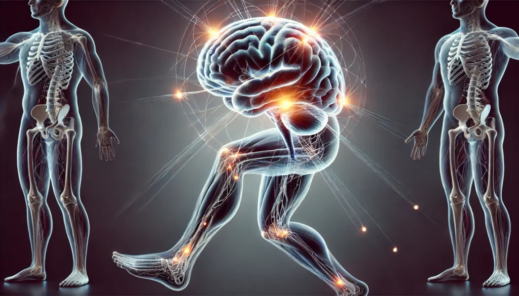
The Cerebellum: The Key to Coordination and Balance
The cerebellum, a structure located at the back of the brain, plays a crucial role in coordinating voluntary muscular movements and maintaining balance. Often referred to as the “little brain,” the cerebellum fine-tunes motor commands from the cerebral cortex to ensure smooth, accurate movements. Without the cerebellum, actions such as picking up a glass or typing on a keyboard would lack precision and fluidity.
One of the cerebellum’s primary functions is to integrate sensory input from the body and adjust motor output accordingly. It receives information from the spinal cord, sensory organs, and cerebral cortex, analyzing the body’s position in space and the required muscular effort for movement. This real-time processing allows for immediate corrections, preventing clumsy or uncoordinated actions. For example, if a person stumbles on an uneven surface, the cerebellum quickly signals adjustments in muscle activity to restore balance.
The role of the cerebellum in balance control is equally significant. It continuously monitors vestibular signals from the inner ear, proprioceptive feedback from muscles and joints, and visual input to maintain stability. This dynamic coordination is essential for standing upright, walking, and engaging in complex activities such as dancing or sports. Damage to the cerebellum, whether due to stroke, injury, or neurodegenerative diseases, can result in ataxia—a condition characterized by impaired coordination, unsteady gait, and difficulty performing fine motor tasks.
The Motor Cortex and Basal Ganglia: Initiating and Refining Movement
While the cerebellum fine-tunes movement, the initiation of voluntary muscular movements originates in the motor cortex. Situated in the frontal lobe, the motor cortex sends signals to the spinal cord and peripheral nerves, directing muscle contractions necessary for movement. Within the motor cortex, the primary motor area (precentral gyrus) plays a key role in executing voluntary actions. Damage to this area can lead to paralysis or motor deficits, underscoring its importance in movement control.
Complementing the motor cortex, the basal ganglia—a collection of deep brain structures—regulate movement intensity and precision. The basal ganglia are responsible for smooth transitions between movements, suppressing unnecessary motor activity while facilitating desired actions. Dysfunction in the basal ganglia is associated with movement disorders such as Parkinson’s disease, characterized by tremors, rigidity, and difficulty initiating movement. This highlights the necessity of a well-functioning motor system for seamless mobility and cognitive-motor integration.
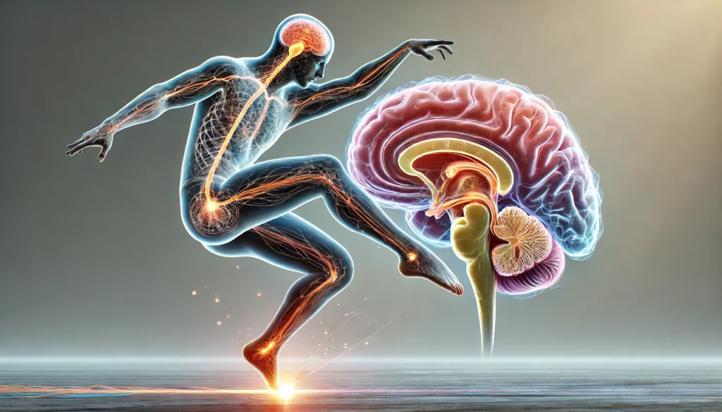
The Role of the Brainstem in Balance and Postural Control
The brainstem, connecting the brain to the spinal cord, is another critical structure involved in balance regulation. Within the brainstem, the vestibular nuclei process signals from the inner ear’s vestibular system, which detects head position and motion. These nuclei communicate with the cerebellum, spinal cord, and ocular motor system to maintain equilibrium. Reflexive postural adjustments, such as shifting weight to prevent falls, depend on these neural circuits.
The reticular formation within the brainstem also contributes to postural control by modulating muscle tone and reflexive movements. Disorders affecting the brainstem, such as strokes or degenerative conditions like multiple system atrophy, can severely impair balance and coordination, increasing the risk of falls and mobility limitations.
Cognitive Health and Movement: The Brain-Body Connection
The ability to coordinate voluntary movements and maintain balance is not solely a physical function—it is deeply intertwined with cognitive health. Research suggests that movement-based activities, such as exercise and dance, enhance neuroplasticity, the brain’s ability to reorganize and adapt. Physical activity stimulates the release of neurotrophic factors, which support neuron survival and connectivity, fostering cognitive resilience against aging-related decline.
Moreover, disruptions in motor coordination and balance can serve as early indicators of neurodegenerative diseases like Alzheimer’s and Parkinson’s. Subtle changes in gait, reaction time, and postural stability may precede cognitive impairment, emphasizing the importance of regular movement assessments in detecting neurological health issues early.
Strategies for Enhancing Brain Function and Mobility
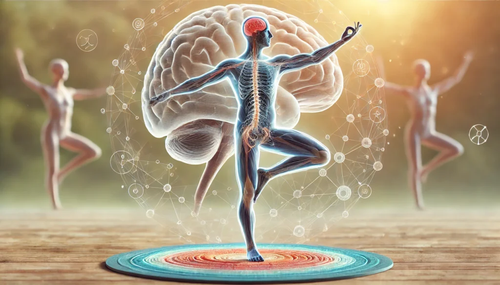
Given the integral relationship between brain function, movement, and balance, adopting lifestyle strategies that support neurological and muscular health is essential. Engaging in regular physical activity, particularly exercises that challenge coordination—such as yoga, Tai Chi, or resistance training—can strengthen neural pathways involved in motor control. Additionally, activities that require dual-tasking, such as playing musical instruments or participating in sports, enhance the brain’s capacity for multitasking and adaptability.
Nutrition also plays a pivotal role in sustaining cognitive-motor health. A diet rich in omega-3 fatty acids, antioxidants, and essential vitamins supports brain function and reduces inflammation, mitigating age-related neural decline. Adequate hydration and sleep further optimize neural signaling, ensuring efficient communication between the brain and muscles.
Frequently Asked Questions (FAQ): Brain Coordination and Balance
1. How does the brain process and refine voluntary movements?
The brain processes voluntary movements through a highly intricate network involving the motor cortex, basal ganglia, and cerebellum. When you decide to move, the motor cortex sends signals to the spinal cord, activating the necessary muscles. However, movement requires more than just initiation; it must be refined to ensure precision and control. The cerebellum plays a critical role in refining these signals by analyzing sensory feedback and adjusting motor commands in real-time. This process ensures that voluntary movements are smooth, coordinated, and adapted to external conditions.
2. What part of the brain coordinates voluntary muscular movements, and how does it ensure accuracy?
The cerebellum is the primary part of the brain that coordinates voluntary muscular movements. It receives input from various sensory systems, including the proprioceptive system, which informs the brain about body position. By integrating this information, the cerebellum fine-tunes motor actions, preventing overshooting or undershooting movements. Additionally, it adapts to repeated tasks through motor learning, meaning that practice leads to improved accuracy over time. This explains why activities like playing an instrument or learning a new sport become more effortless with repetition.
3. Which part of the brain controls balance, and what mechanisms are involved?
The brainstem and cerebellum are the primary regions responsible for balance control. The brainstem houses the vestibular nuclei, which process signals from the inner ear’s vestibular system to detect head position and motion. The cerebellum interprets these signals alongside visual and proprioceptive input, allowing the body to make rapid postural adjustments. The integration of these signals ensures that balance is maintained even when moving or standing on uneven surfaces. Damage to these areas can result in dizziness, unsteady gait, or difficulty maintaining equilibrium.
4. How does aging affect the part of the brain for balance?
Aging naturally impacts the part of the brain for balance, particularly the cerebellum and brainstem. Neural connections may weaken, reducing the efficiency of sensory processing and motor coordination. Additionally, age-related changes in the vestibular system can lead to impaired spatial awareness and slower postural reflexes. This decline often manifests as an increased risk of falls, making balance training exercises such as Tai Chi or yoga essential for maintaining stability. Strengthening core muscles and engaging in proprioceptive training can also help counteract balance deterioration in older adults.
5. How do neurological disorders affect voluntary movements?
Neurological disorders such as Parkinson’s disease and multiple sclerosis directly impact the brain’s ability to coordinate voluntary muscular movements. In Parkinson’s disease, the basal ganglia experience dopamine depletion, leading to tremors, rigidity, and difficulty initiating movement. Multiple sclerosis affects the myelin sheath that insulates nerve fibers, causing disruptions in signal transmission and leading to muscle weakness or coordination issues. Early intervention with physical therapy and medication can help manage symptoms and improve motor control.
6. Can brain exercises improve the part of the brain for balance?
Yes, brain exercises can enhance the part of the brain for balance by strengthening neural connections and improving coordination. Activities that challenge cognitive-motor integration, such as dance, balance board exercises, and dual-tasking challenges, stimulate the cerebellum and brainstem. Meditation and mindfulness training can also improve body awareness, allowing for better balance control. Engaging in these exercises regularly can reduce fall risk and improve overall stability, particularly in aging individuals or those recovering from injury.
7. What lifestyle habits support the brain’s coordination of voluntary movements?
Healthy lifestyle habits such as proper nutrition, regular exercise, and quality sleep are crucial for maintaining the brain’s ability to coordinate voluntary muscular movements. Omega-3 fatty acids, found in fish and nuts, support brain cell communication, while antioxidants combat oxidative stress that can impair neural function. Strength training and aerobic exercise promote neuroplasticity, keeping motor pathways efficient. Additionally, getting adequate rest allows the brain to consolidate motor learning, reinforcing movement patterns developed during waking hours.
8. How does the vestibular system interact with the brain to maintain balance?
The vestibular system, located in the inner ear, plays a vital role in balance by detecting head movement and spatial orientation. It sends signals to the vestibular nuclei in the brainstem, which then communicate with the cerebellum to make postural adjustments. This interaction ensures that head and body movements are synchronized, preventing dizziness and disorientation. When vestibular function is impaired due to injury or illness, individuals may experience vertigo or difficulty maintaining stability. Vestibular rehabilitation therapy can help retrain the brain to process balance-related signals effectively.
9. What role does proprioception play in maintaining stability?
Proprioception, or the body’s ability to sense its position in space, is a crucial component of balance and movement coordination. Sensory receptors in the muscles, joints, and tendons relay information to the cerebellum and brainstem, helping the brain adjust posture and movement accordingly. Without proprioception, even simple tasks like walking would become difficult, as the body would struggle to detect changes in position. Strengthening proprioceptive awareness through exercises like single-leg stances, agility drills, and coordination training can enhance stability and prevent falls.
10. How do head injuries impact the brain’s ability to control balance and voluntary movement?
Head injuries, particularly concussions and traumatic brain injuries (TBIs), can severely affect the brain’s ability to control balance and voluntary movement. Damage to the cerebellum, motor cortex, or vestibular system can result in coordination deficits, dizziness, and impaired spatial awareness. Even mild TBIs can lead to lingering balance problems and delayed reaction times. Rehabilitation programs that focus on neuroplasticity, including balance retraining, vestibular therapy, and cognitive exercises, can help restore function. Protecting the brain through helmet use, fall prevention strategies, and safe physical activities is essential for minimizing injury risk.
Conclusion: Preserving Brain Health for Lifelong Mobility
Understanding which part of the brain coordinates voluntary muscular movements and which part of the brain controls balance provides valuable insights into maintaining cognitive health and longevity. The cerebellum, motor cortex, basal ganglia, and brainstem work in harmony to facilitate movement and stability, emphasizing the brain’s intricate role in mobility. By prioritizing physical activity, balanced nutrition, and cognitive engagement, individuals can preserve their brain’s ability to regulate movement, reducing the risk of neurodegenerative conditions and enhancing overall well-being. As research continues to uncover the interconnectedness of brain function and motor control, integrating movement-focused strategies into daily life remains a key pillar of longevity and cognitive vitality.
brain function and movement, motor control in the brain, neurological coordination, cerebellum and motor skills, balance disorders treatment, brain health and mobility, cognitive function and movement, nervous system and coordination, proprioception and stability, neuroplasticity and motor learning, vestibular system function, postural control mechanisms, aging and motor skills, brainstem role in balance, movement disorders research, Parkinson’s disease and coordination, strengthening balance through exercise, neurological rehabilitation techniques, sensory integration and balance, motor cortex and voluntary action
Further Reading:
Which Parts of the Brain Are Responsible for Balance and Equilibrium?
Movement: How the Brain Communicates with the World
Understanding Cerebellum Function: The Brain’s Control Center
Disclaimer
The information contained in this article is provided for general informational purposes only and is not intended to serve as medical, legal, or professional advice. While Health11News strives to present accurate, up-to-date, and reliable content, no warranty or guarantee, expressed or implied, is made regarding the completeness, accuracy, or adequacy of the information provided. Readers are strongly advised to seek the guidance of a qualified healthcare provider or other relevant professionals before acting on any information contained in this article. Health11News, its authors, editors, and contributors expressly disclaim any liability for any damages, losses, or consequences arising directly or indirectly from the use, interpretation, or reliance on any information presented herein. The views and opinions expressed in this article are those of the author(s) and do not necessarily reflect the official policies or positions of Health11News
