

Introduction: Decoding the Brain’s Complex Landscape
The human brain is a marvel of evolution, boasting an intricate structure that reflects its profound cognitive capabilities. Among its most visually distinctive features are the folds and grooves that mark its surface, a pattern so unique that even a cursory glance at an anatomical diagram reveals their complex arrangement. But what are the folds of the brain called, and why do they exist in the first place? These grooves, also known as sulci, and their raised counterparts, called gyri, are far more than just surface-level wrinkles. They are critical architectural features that enable the brain to maximize its cortical area within the confined space of the skull. By folding in on itself, the brain can support a larger volume of gray matter, which is directly tied to higher cognitive function. This fundamental design principle plays a crucial role in learning, memory, emotional regulation, sensory perception, and overall mental health. Understanding what the wrinkles on the brain are called and their function is not only an academic exercise but also a foundational step toward appreciating how structural variations can affect mental well-being and neurological health. From developmental neuroscience to the study of mental disorders like schizophrenia and depression, the morphology of brain folds offers a window into the inner workings of our most vital organ.
You may also like: Boost Brain Power Naturally: Evidence-Based Cognitive Training Activities and Memory Exercises That Support Long-Term Mental Health
The Terminology and Anatomy Behind Brain Folds
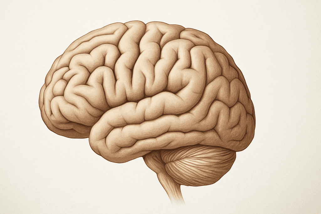
To fully grasp the meaning and function of these brain structures, we must first explore their proper anatomical terminology. The folds on the brain are called gyri (singular: gyrus), while the indentations or grooves that separate them are known as sulci (singular: sulcus). This folded architecture is most prominently seen in the cerebral cortex, the outermost layer of the brain, which is responsible for higher-order functions such as thought, language, decision-making, and sensory processing. These folds increase the surface area of the cortex significantly, allowing for a greater number of neurons to be packed into a limited space. In fact, if the human brain were to be unfolded, its cortical surface area would span approximately 2,500 square centimeters, or nearly the size of a large pillowcase. This expansive surface area is essential for accommodating the approximately 86 billion neurons that populate the brain, forming complex networks responsible for every sensation, movement, and thought we experience. So when we ask what the folds of the brain are called, we’re not merely identifying anatomical landmarks—we’re acknowledging a sophisticated system designed to optimize our neurological capacity. Furthermore, individual sulci and gyri often have specific names and are associated with distinct brain regions and functions. For example, the precentral gyrus plays a key role in motor control, while the postcentral gyrus is critical for processing somatosensory information. The central sulcus, which separates these two regions, serves as an important boundary marker between different functional areas of the brain.
Evolutionary Significance of Brain Wrinkles
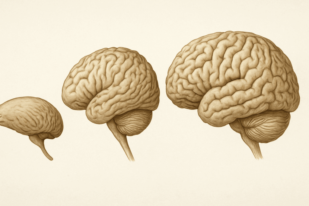
The reason behind the brain’s wrinkled appearance lies in the evolutionary pressures that have shaped the human species. Compared to our primate relatives, humans possess a significantly more folded cerebral cortex—a feature known as gyrencephaly. This increased folding is believed to be directly linked to our advanced cognitive abilities, such as abstract reasoning, problem-solving, and social interaction. In evolutionary biology, the degree of cortical folding is often used as a marker of brain complexity and intelligence. Species with smoother brains, like rodents, exhibit less complex behaviors, while those with highly convoluted cortices, such as dolphins and great apes, display sophisticated communication skills and social structures. Understanding what the wrinkles on the brain are called and why they evolved provides key insights into our neurological uniqueness. The folds enable the brain to house more neurons and support intricate neural circuits that underpin complex behavior. These adaptations allowed early humans to survive and thrive in diverse environments by enhancing their ability to learn, adapt, and communicate. In addition to their cognitive benefits, the brain’s wrinkles also contribute to its metabolic efficiency. By allowing more cortical surface area to be packed into a small volume, the brain can maintain shorter average distances between neurons, facilitating faster signal transmission. This efficient wiring conserves energy while preserving rapid and reliable communication across different brain regions. Thus, the evolution of gyri and sulci is not merely a structural adaptation—it’s a testament to the brain’s ability to balance performance with efficiency, a key hallmark of intelligent biological design.
How Folding Affects Brain Function
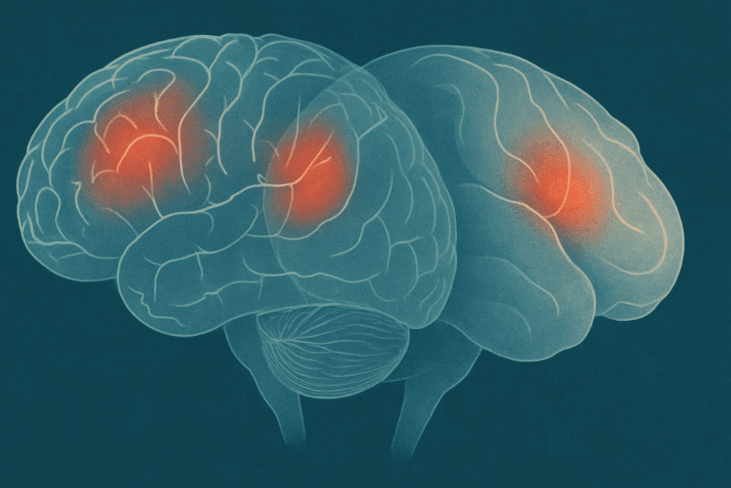
When considering what the folds of the brain are called and why they matter, it’s essential to understand how they influence the brain’s functional organization. The folding patterns create boundaries that often correspond to different brain regions, each responsible for specific cognitive, emotional, or sensory tasks. These gyri and sulci guide the segregation and integration of neural processes, allowing specialized areas to carry out distinct functions while maintaining communication with other regions. For instance, the temporal lobe, tucked beneath the lateral sulcus, plays a vital role in auditory processing and language comprehension. Meanwhile, the occipital lobe, bordered by the parieto-occipital sulcus, is the brain’s visual processing center. The structural delineation provided by these folds ensures that functional domains are both spatially organized and interconnected. Alterations in these folding patterns, whether due to developmental anomalies or neurological disease, can significantly impact cognitive function. Conditions such as polymicrogyria and lissencephaly—where the brain exhibits too many or too few folds—are associated with intellectual disabilities, seizures, and motor dysfunction. Even subtle deviations in cortical folding have been observed in individuals with autism, schizophrenia, and major depressive disorder. This highlights the profound connection between brain morphology and mental health. By studying what the wrinkles on the brain are called and how they form, neuroscientists can better understand the structural biomarkers that underlie various psychiatric and neurological conditions. Moreover, advances in neuroimaging techniques now allow researchers to map individual variations in brain folding with remarkable precision, opening new avenues for personalized medicine and early diagnosis.
Brain Development and the Formation of Gyri and Sulci
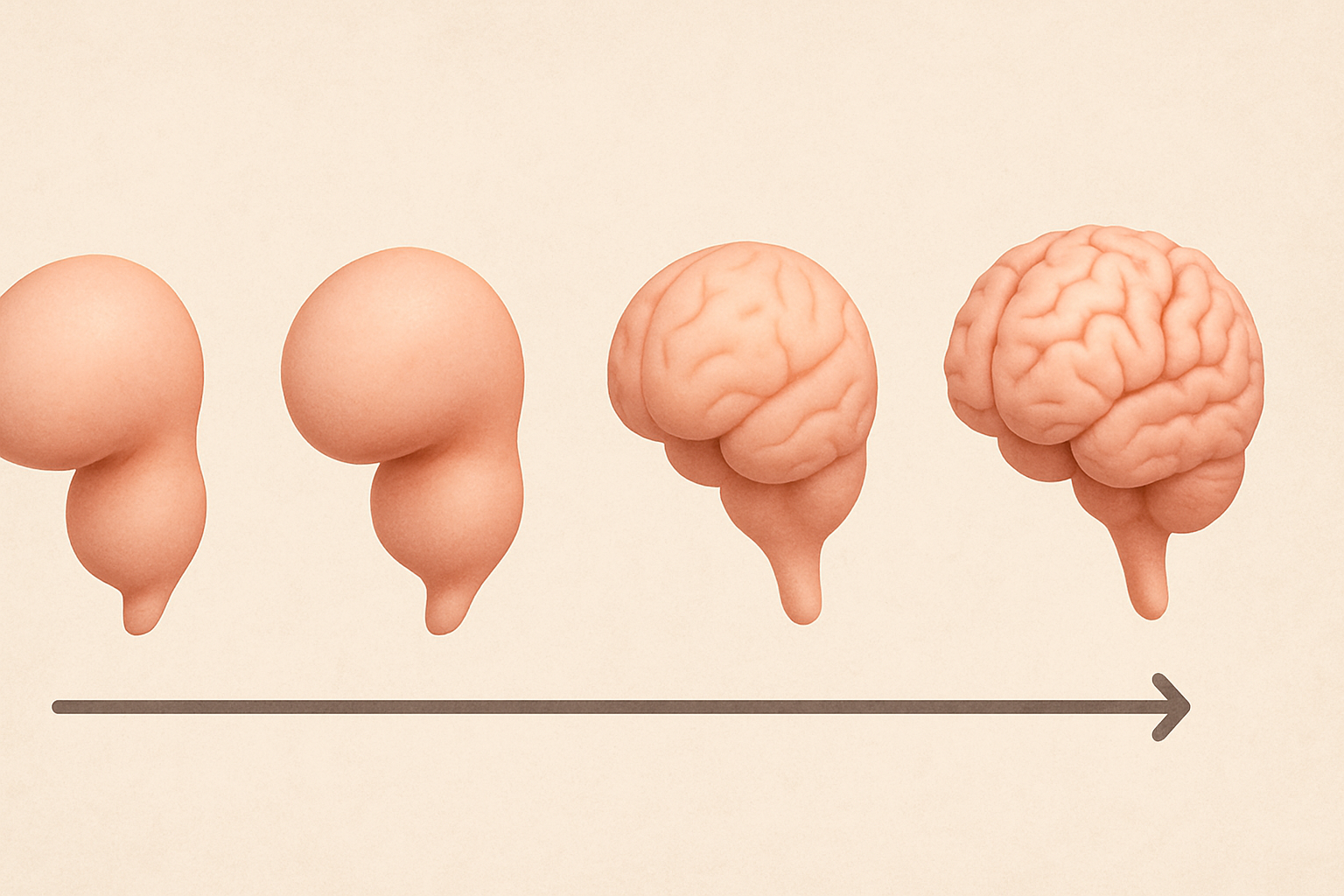
The process of brain folding begins during fetal development and continues into early childhood, following a highly orchestrated timeline that reflects genetic and environmental influences. Around the 20th week of gestation, the previously smooth fetal brain starts to develop shallow grooves and ridges that gradually deepen and proliferate. This process, known as gyrification, transforms the cerebral cortex into the characteristic wrinkled surface we recognize in mature brains. The primary sulci, such as the central and lateral sulci, form first and establish the basic layout of cortical regions. Secondary and tertiary folds develop later, reflecting finer functional specialization and increasing interconnectivity. Genetic mutations or environmental stressors that disrupt this timeline can lead to atypical folding patterns, which may underlie a range of neurodevelopmental disorders. For example, abnormal gyrification has been implicated in schizophrenia and bipolar disorder, suggesting that even subtle changes in brain surface anatomy can have profound effects on cognition and mood. Understanding what the folds of the brain are called becomes especially significant when examining developmental trajectories. These folds not only represent anatomical structures but also markers of healthy brain maturation. Moreover, the rate and pattern of folding can vary across individuals, contributing to unique cognitive strengths and vulnerabilities. Recent studies using MRI technology have revealed that individuals with higher cortical complexity—more intricate folding patterns—tend to perform better on tests of memory, attention, and executive function. Thus, the morphology of gyri and sulci serves as a dynamic reflection of both biological inheritance and life experience, bridging the gap between brain structure and mental performance.
Mental Health and the Structure of the Cerebral Cortex
Exploring what the wrinkles on the brain are called inevitably leads us to their profound implications for mental health. The morphology of the cerebral cortex has been increasingly recognized as a biomarker for various psychiatric conditions. In depression, for instance, researchers have observed altered gyrification in regions like the prefrontal cortex—areas associated with mood regulation and executive function. Similarly, individuals with anxiety disorders often exhibit structural differences in the insular cortex and anterior cingulate gyrus, regions that mediate emotional awareness and stress response. These findings suggest that brain folding patterns are not just passive byproducts of development but actively shape our emotional and cognitive landscape. In disorders like schizophrenia, aberrant cortical folding has been linked to disruptions in neural connectivity, which may account for symptoms such as hallucinations, disorganized thinking, and impaired working memory. The condition is often associated with reduced gyrification in the frontal and temporal lobes, impairing the brain’s ability to process complex information efficiently. Autism spectrum disorder also presents with unique gyrification signatures, often involving increased folding in some areas and decreased folding in others, reflecting atypical patterns of brain network organization. By understanding what the folds of the brain are called and how they relate to specific functional areas, clinicians and researchers can better interpret the neuroanatomical correlates of mental illness. Importantly, these insights are paving the way for earlier detection and more targeted interventions. As our understanding of the brain’s surface morphology deepens, so does our ability to devise treatment plans that are not only symptom-based but grounded in structural and functional neuroscience.
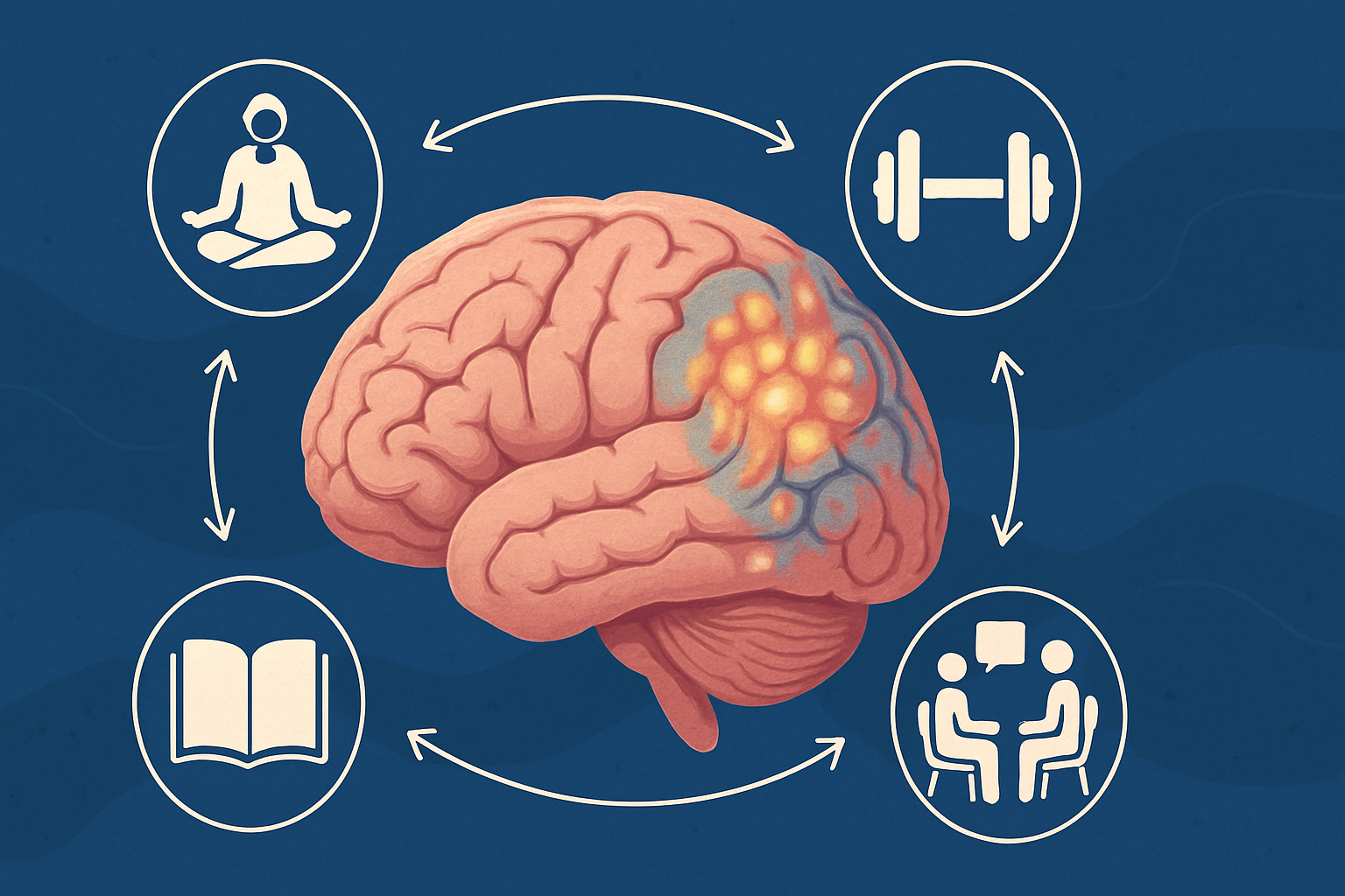
Brain Plasticity and the Changing Landscape of Cortical Folding
One of the most remarkable features of the human brain is its plasticity—the ability to adapt and reorganize in response to experience, learning, and injury. While major folding patterns are largely established during development, emerging research suggests that the brain’s surface can continue to evolve throughout life. This is particularly evident in the context of neuroplastic changes induced by cognitive training, therapy, or recovery from injury. Studies have shown that individuals who engage in activities that challenge the brain—such as learning a new language, playing a musical instrument, or practicing mindfulness meditation—exhibit changes in cortical thickness and even subtle alterations in gyrification. These adaptations are especially prominent in regions involved in attention, memory, and emotion regulation. In this context, understanding what the wrinkles on the brain are called takes on a renewed significance, as these structures are not static but responsive to the rhythms of life. Moreover, neuroplasticity holds promise for mental health interventions. For example, cognitive-behavioral therapy (CBT) has been shown to induce structural changes in the prefrontal cortex of individuals with depression and anxiety, potentially reversing maladaptive folding patterns associated with these disorders. Similarly, physical exercise and enriched environments have been linked to increased cortical complexity in animal models, suggesting that lifestyle choices can influence brain architecture. The emerging field of neuroarchitecture—how the brain’s shape affects function and vice versa—is shedding new light on the potential for recovery and enhancement. Thus, the question of what the folds of the brain are called extends beyond static anatomy; it touches on the dynamic interplay between structure, function, and experience, affirming the brain’s remarkable ability to reshape itself in pursuit of wellness.
Frequently Asked Questions: Understanding the Brain’s Wrinkles and Folds
I. How do the brain’s folds influence individual personality traits?
While research into the relationship between brain anatomy and personality is ongoing, emerging evidence suggests that the patterns of folds—known as gyri and sulci—can influence behavioral tendencies. These structures impact how neural networks are formed and how efficiently they communicate. Subtle variations in the folding of the prefrontal cortex, for example, have been associated with differences in impulse control, empathy, and decision-making. So, when considering what the folds of the brain are called, it’s worth exploring how these anatomical features may support the neural basis of personality. Although genes play a major role, experiences and environments that shape folding patterns during development could also contribute to personality differences across individuals.
II. Can traumatic brain injury alter the wrinkles on the brain?
Yes, traumatic brain injuries (TBI) can cause changes in the brain’s surface, particularly in severe or repeated cases. Damage from TBI can result in swelling, loss of gray matter, or cortical atrophy, all of which may affect the structure of gyri and sulci. In some cases, these changes disrupt the intricate folding patterns, leading to impaired cognitive or emotional functioning. When clinicians assess long-term impacts of TBI, imaging often reveals abnormalities in areas where what are the wrinkles on the brain called are no longer as well defined. Understanding these structural consequences helps healthcare providers better predict functional impairments and guide rehabilitation strategies.
III. Are there gender differences in brain folding patterns?
Studies have shown that while overall brain structure is more similar than different between genders, there can be subtle variations in folding patterns. Women often exhibit a higher degree of cortical gyrification in certain brain regions, which may relate to enhanced interconnectivity and multitasking abilities. These findings don’t imply superiority but rather reflect distinct architectural variations shaped by both biology and social experience. When researchers explore what the folds of the brain are called, they sometimes discover gender-linked folding differences that correlate with unique cognitive strengths. However, it’s crucial to interpret these patterns as part of a broader mosaic of brain function rather than rigid distinctions.
IV. How do folding patterns change as we age?
As the brain ages, natural structural changes occur, including subtle flattening of the gyri and widening of the sulci. These shifts can reflect a loss of cortical volume and reduced synaptic density, though they vary greatly between individuals. Age-related folding changes are more pronounced in areas like the frontal lobe, which is associated with memory and executive function. When imaging reveals diminished definition in what are the wrinkles on the brain called, it often correlates with mild cognitive decline. That said, maintaining an active lifestyle and engaging in mentally stimulating activities can help preserve the integrity of these structures well into older adulthood.
V. Is there a connection between neurodivergence and brain wrinkles?
Yes, research into conditions like autism spectrum disorder (ASD), ADHD, and dyslexia has uncovered distinctive differences in cortical folding patterns. Individuals with ASD, for instance, may show increased gyrification in certain brain regions, reflecting atypical developmental trajectories. These differences affect how various areas of the brain communicate, which can influence behavior, learning style, and sensory processing. Recognizing what the wrinkles on the brain are called can be helpful for clinicians and researchers trying to identify neuroanatomical markers of neurodivergence. While folding patterns alone don’t diagnose a condition, they can offer valuable insight into the brain’s unique organization in neurodiverse populations.
VI. Can lifestyle factors influence the structure of brain folds?
Surprisingly, lifestyle choices such as exercise, diet, stress management, and intellectual engagement can affect the brain’s structure over time. Regular aerobic activity has been shown to preserve gray matter and may even slow the degradation of gyri and sulci in aging populations. Chronic stress, on the other hand, can negatively affect regions where what are the wrinkles on the brain called are most complex, such as the hippocampus. Engaging in cognitively challenging activities—like learning a language or playing strategy games—can also promote cortical plasticity. Although folding patterns are largely set in early development, these lifestyle factors can help maintain brain health and adaptability.
VII. Do artificial intelligence models help map brain folds more accurately?
Yes, advanced artificial intelligence (AI) tools have revolutionized how we analyze brain imaging data. Modern algorithms can identify subtle variations in cortical folding across large datasets, enabling more precise diagnosis of neurological conditions. AI is also helping scientists understand what the folds of the brain are called in new ways—by linking structural complexity to functional outcomes with greater accuracy. These models can detect patterns the human eye might miss and assist in creating personalized neurodevelopmental profiles. In clinical settings, AI-driven imaging may one day support early detection of mental health disorders by flagging atypical folding patterns linked to cognitive risk factors.
VIII. How are folding patterns different in highly creative individuals?
Recent neuroscience research suggests that individuals with exceptional creativity may show unique patterns of cortical folding, especially in regions related to imagination and abstract thinking. Enhanced connectivity between the default mode network and the executive control network is sometimes supported by more intricate folding in the frontal and temporal lobes. While it’s too early to make broad generalizations, studies are beginning to explore how variations in what the wrinkles on the brain are called could support divergent thinking and innovation. Creativity likely emerges from a combination of structural, functional, and environmental factors, but the physical architecture of the brain plays a compelling role. These insights challenge the idea that genius is purely innate, suggesting it may also be structurally supported.
IX. Could abnormal brain wrinkles be used to predict future mental health issues?
Researchers are investigating whether atypical folding patterns could serve as early biomarkers for mental health risks. For example, reduced gyrification in the anterior cingulate cortex has been linked to vulnerability to depression and anxiety. Identifying abnormalities in what are the folds of the brain are called may help clinicians monitor individuals at higher risk for developing conditions like schizophrenia, even before symptoms emerge. This predictive capability could allow for preventive mental health strategies, personalized therapy plans, and earlier intervention. However, more longitudinal studies are needed to validate these associations across diverse populations and determine their practical applications in clinical psychiatry.
X. Are there cultural or educational factors that affect brain folding patterns?
While genetics and biology are primary drivers of brain structure, cultural and educational environments also play an influential role. Exposure to multilingual education, music training, and even philosophical reasoning can shape the development of brain areas associated with learning and abstraction. These experiences may subtly influence how and where the folds of the brain develop enhanced complexity or density. Though cultural effects on gyrification are still being studied, it’s increasingly clear that the brain is responsive to environmental enrichment. This supports the broader view that the brain’s architecture, while biologically grounded, remains sensitive to the social and intellectual context in which a person lives and learns.
Conclusion: Why Understanding Brain Wrinkles Matters for Cognitive and Mental Health
In the quest to understand human cognition and mental health, few questions are as deceptively simple yet profoundly revealing as: What are the folds of the brain called? These gyri and sulci, the wrinkles on the brain that give it its signature appearance, are much more than topographical curiosities. They are vital components of a highly efficient and intricately organized system designed to support the full spectrum of human experience—from memory and emotion to decision-making and social connection. The patterns of these folds influence not only how we think and feel but also how we respond to stress, adapt to change, and recover from mental illness. By exploring what the wrinkles on the brain are called and how they function, we gain invaluable insights into the relationship between structure and well-being. This knowledge empowers researchers, clinicians, and individuals alike to better understand the roots of mental health disorders, track developmental progress, and even harness the brain’s plasticity for personal growth and healing. As we continue to advance our tools for imaging and analyzing the brain, the once-mysterious folds on its surface are becoming powerful markers of neurological integrity and cognitive potential. In essence, the wrinkles of the brain are not merely signs of biological engineering—they are the very scaffolding of our minds, shaping the thoughts, emotions, and behaviors that define our lives. Understanding their function is not just a matter of scientific curiosity; it is a cornerstone of modern mental health and wellness.
Further Reading:
Some People’s Brains Are Wrinklier Than Others, And Now We Know Why
