

Understanding the Complexity of the Human Brain
The human brain is the most intricate and enigmatic organ in the body. As the control center for all thought, behavior, and physiological regulation, it is responsible for our ability to speak, think, feel, and act. Anatomically, the brain consists of multiple regions and lobes, each responsible for distinct functions, yet working in coordination through billions of synaptic connections. Advances in medical imaging and neuroscience have made it possible to visualize and study these structures in remarkable detail. High-resolution brain structure diagrams and labeled human brain diagrams have transformed how we understand cognition, behavior, and neurological disorders.
A detailed picture of the brain labeled correctly can help medical professionals, students, and curious learners identify regions such as the prefrontal cortex, cerebellum, brainstem, and limbic system with clarity. These pictures of brain anatomy are more than academic tools—they serve as foundational visual references in diagnosing traumatic brain injuries, neurodegenerative diseases, and developmental abnormalities. Understanding what lies inside the brain side view labeled diagrams reveals the spatial orientation of each structure in real-world clinical contexts. With each labeled human brain diagram, there is a deepening of our comprehension of the organ that defines our humanity.
Whether exploring a cerebral picture for educational purposes or reviewing a brain parts chart for clinical precision, this guide will explore each critical region of the brain with in-depth descriptions. From the forebrain diagram that details executive functions to the top view of brain-labeled images that display interlobular boundaries, every visual aid tells a story of form and function. These brain part images are essential tools for anyone seeking to label the brain structures accurately and appreciate the marvel of human anatomy.
You may also like: Boost Brain Power Naturally: Evidence-Based Cognitive Training Activities and Memory Exercises That Support Long-Term Mental Health
Mapping the Lobes: A Visual and Functional Overview
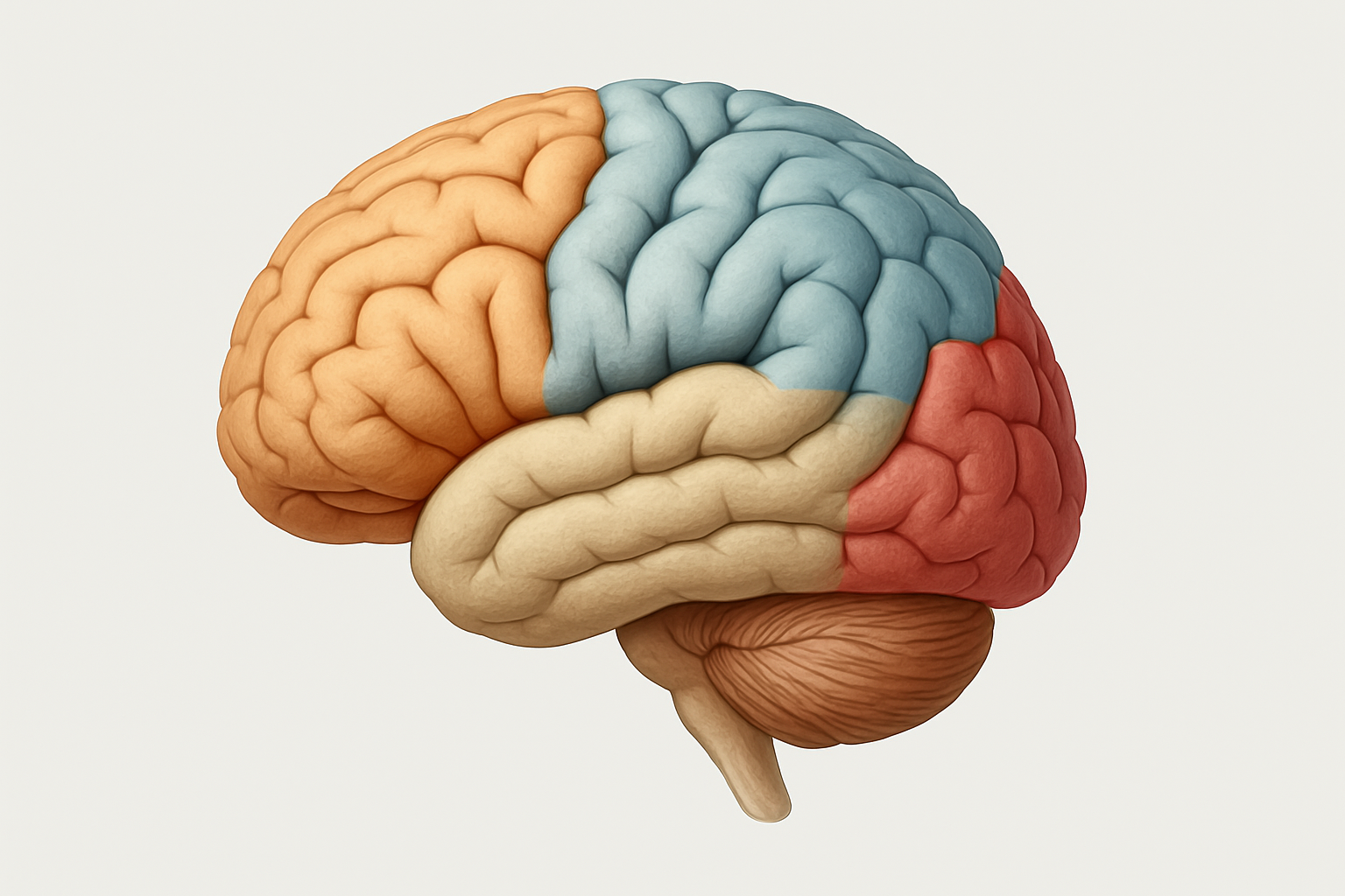
One of the most compelling features of the brain is its division into four major lobes: frontal, parietal, temporal, and occipital. Each lobe is associated with unique responsibilities, and understanding them is critical for interpreting a picture of brain lobes or analyzing a labeled human brain diagram. The frontal lobe, located at the front of the brain, governs higher-order functions such as decision-making, emotional regulation, and voluntary motor activity. Its importance is underscored in brain structure diagrams, where it appears as the center of executive control. Damage to this area can result in changes to personality, difficulty concentrating, or impaired movement.
The parietal lobe lies behind the frontal lobe and is primarily responsible for processing sensory information such as touch, temperature, and pain. When reviewing a picture of the brain labeled for sensory interpretation, this region is often shaded or annotated for its role in somatosensory integration. This lobe also plays a role in spatial awareness and navigation, making it crucial for understanding how we relate physically to our environment. Temporal lobes, found beneath the parietal lobes on each side of the brain, are key to auditory processing, language comprehension, and memory formation. Brain parts images that include the temporal lobe often highlight its deep involvement with the hippocampus and amygdala—structures integral to emotional and memory-related functions.
Finally, the occipital lobe, positioned at the rear of the brain, is the visual processing hub. If one were to look at a top view of the brain labeled for visual input, this lobe would dominate the rear section, with pathways connecting it to the eyes via the optic nerves. Understanding this layout is particularly vital when analyzing pictures of brain anatomy that correlate vision with specific neurological pathways. By exploring each lobe in relation to its responsibilities, students and clinicians alike can use brain parts charts and labeled human brain diagrams to navigate neurological assessments with greater confidence and clarity.
The Forebrain: Executive Control and Emotional Processing
The forebrain represents the most advanced portion of the human brain and is home to the cerebral cortex and subcortical structures such as the thalamus, hypothalamus, and basal ganglia. When examining a forebrain diagram or a cerebral picture, one is essentially looking at the command center of cognition, emotion, and voluntary behavior. The cerebral cortex, the outermost layer of the forebrain, is involved in everything from reasoning and abstract thought to sensory perception and motor control. This area is often prominently featured in a labeled human brain diagram due to its significance in virtually all aspects of conscious life.
A well-designed picture of the human brain labeled with detailed cortical and subcortical areas can help explain the nuanced functions governed by the forebrain. For example, the prefrontal cortex within the frontal lobe is responsible for planning and decision-making, while the limbic system, which includes the amygdala and hippocampus, is central to emotion and memory. These connections underscore how brain structure diagrams are vital for understanding behavioral and psychological phenomena. When viewing images of brain parts in clinical neuropsychology, the focus often centers on how damage or dysfunction in the forebrain correlates with psychiatric conditions such as depression, anxiety, or schizophrenia.
In addition to its psychological influence, the forebrain also plays a major role in regulating physiological functions. The hypothalamus, for instance, helps control hunger, thirst, sleep, and hormonal processes through its link to the endocrine system. Labeling the brain structures in this region provides clarity in understanding conditions such as insomnia, endocrine disorders, and even metabolic syndromes. A comprehensive pic of brain parts showing this region enables researchers and clinicians to pinpoint dysfunctions and intervene with targeted treatments. The forebrain, as represented in detailed labeled diagrams and cerebral pictures, remains at the forefront of neuroscience due to its unparalleled complexity and functional breadth.
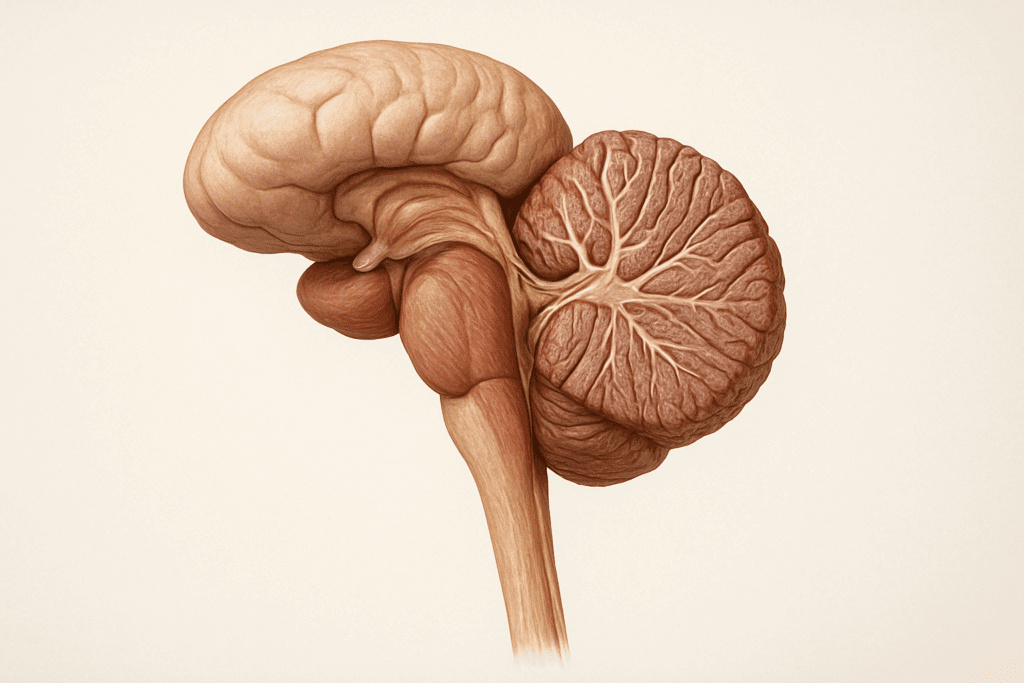
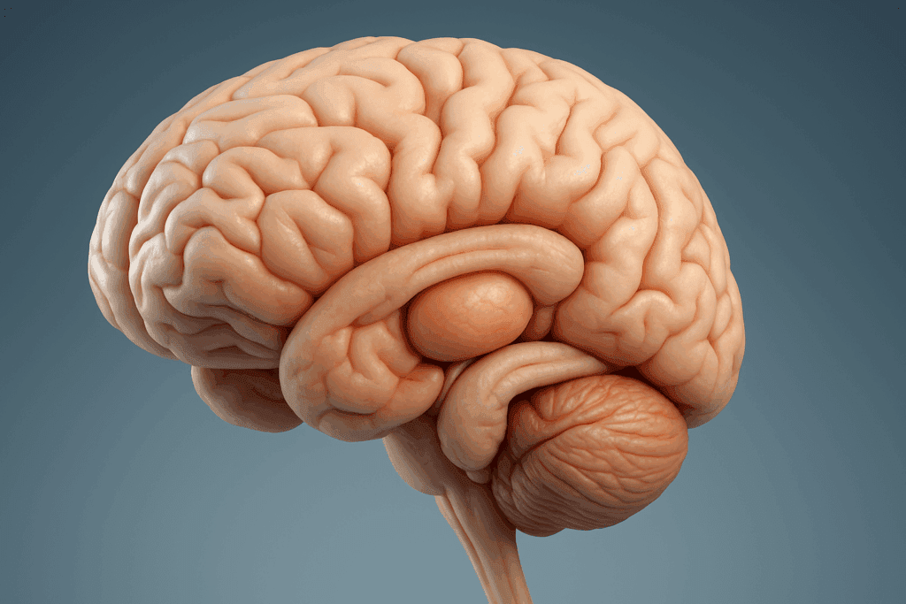
The Brainstem and Cerebellum: Life Support and Motor Coordination
While the forebrain often steals the spotlight due to its role in higher cognition, the brainstem and cerebellum are no less vital. These structures form the core of the hindbrain and midbrain, regions responsible for sustaining life and regulating essential autonomic functions. A picture of the brain parts that includes the brainstem reveals its tripartite division into the midbrain, pons, and medulla oblongata. Each component manages different physiological processes such as breathing, heart rate, digestion, and sleep-wake cycles. A labeled human brain diagram that clearly marks the brainstem is crucial for medical students learning about neurological reflexes and vital control centers.
The cerebellum, located just beneath the occipital lobe and behind the brainstem, is the master controller of balance, posture, and fine motor coordination. It receives input from the sensory systems, spinal cord, and other parts of the brain to fine-tune movement and ensure smooth execution. When examining brain parts charts or images, the cerebellum often appears as a distinct, leaf-like structure due to its folded surface, known as folia. This anatomical detail is critical when assessing cerebellar function through neuroimaging, especially in cases of stroke, trauma, or neurodegenerative disorders such as ataxia.
A detailed inside brain side view label can help learners and clinicians alike appreciate the anatomical continuity between the cerebrum, cerebellum, and brainstem. This integration is essential for understanding how the brain functions as a holistic system rather than a collection of isolated parts. In emergency medicine and neurology, the rapid interpretation of pictures of brain anatomy that include these regions can make the difference between timely intervention and irreversible damage. The structural integrity and interaction between the cerebellum and brainstem ensure the seamless coordination of voluntary actions with involuntary physiological processes, underscoring their significance in maintaining life and movement.
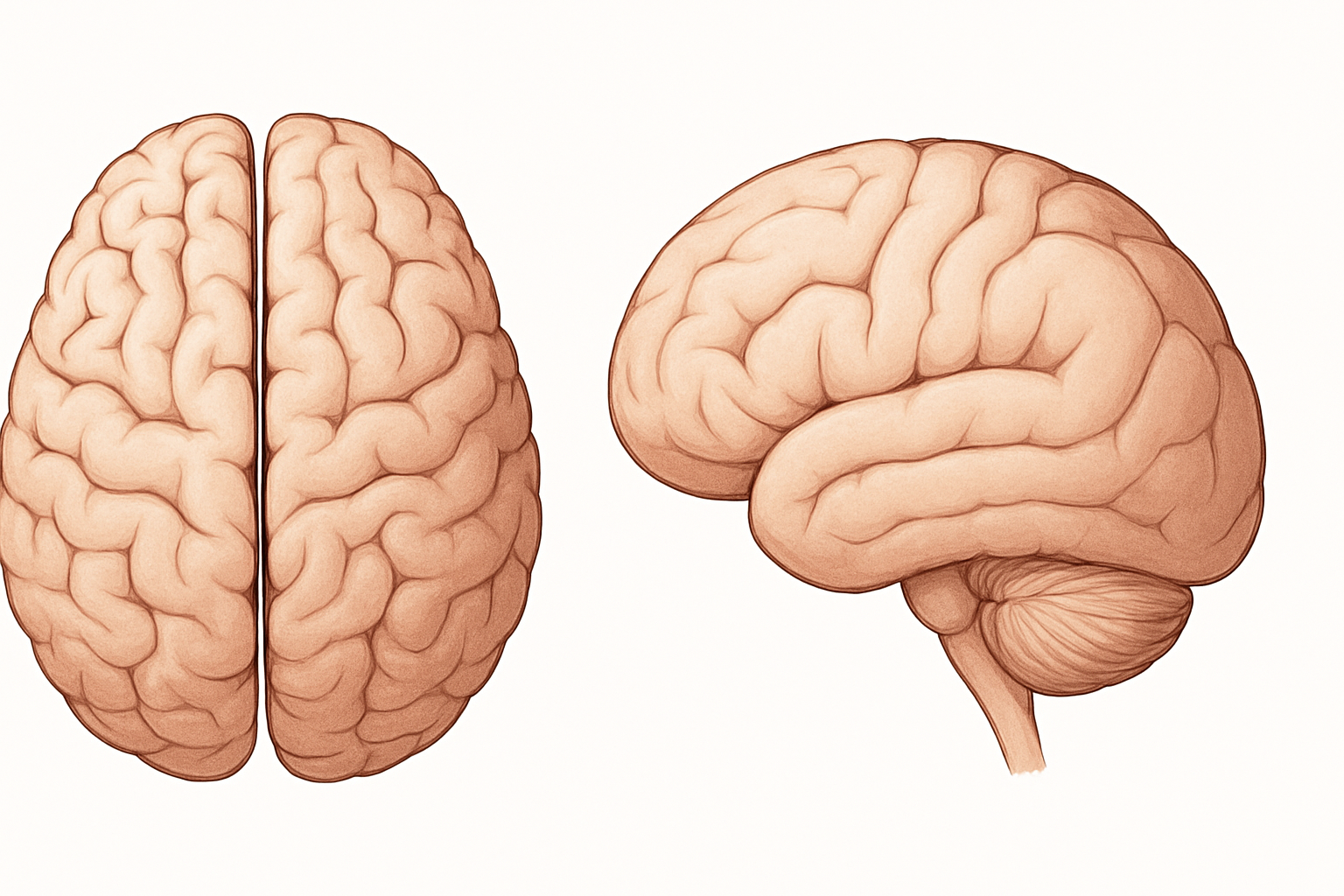
Lateral and Top Views: Spatial Orientation and Structural Insight
Understanding the spatial layout of the brain is essential for both clinical and academic contexts. A top view of the brain labeled offers a bird’s-eye perspective, often used to differentiate between the two cerebral hemispheres and identify interlobular borders. This perspective is particularly helpful when comparing functional hemispheric dominance, such as language being typically lateralized to the left hemisphere. Brain structure diagrams viewed from the top also provide an overview of the longitudinal fissure that separates the hemispheres and the corpus callosum that connects them. This orientation aids in assessing callosal syndromes and interhemispheric disconnection.
Meanwhile, an inside brain side view provides insight into the medial structures such as the cingulate gyrus, corpus callosum, and ventricular system. These diagrams are especially useful in neuroimaging, where sagittal slices of the brain reveal white matter tracts and deep nuclei. A picture of brain lobes from the side can illustrate how each lobe folds into the next and how sulci and gyri enhance cortical surface area. Such visual information is essential for planning surgical procedures or diagnosing lesions visible on MRI and CT scans. For example, in epilepsy treatment, lateral brain imaging is often used to locate foci for potential resection.
When labeling the brain structures in these views, accuracy is paramount. A mislabeled diagram could lead to misconceptions in learning or even diagnostic errors in practice. Therefore, brain parts charts and labeled human brain diagrams used in medical settings are typically peer-reviewed and standardized. These images not only serve as study aids but also as diagnostic tools that bridge the gap between structural knowledge and functional understanding. Whether you’re a medical student, neurologist, or educator, using a comprehensive picture of the brain parts with proper labels enhances your ability to interpret, explain, and act upon the information that the brain reveals.
Frequently Asked Questions (FAQ) About the Human Brain and Its Labeled Structures
1. What are some advanced ways that researchers use a picture of brain labeled in neurological diagnostics?
A picture of brain labeled goes far beyond classroom anatomy—it plays a central role in advanced diagnostics such as identifying the precise location of tumors, tracking the progression of degenerative diseases like multiple sclerosis, and evaluating the impact of traumatic injuries. Researchers use these labeled brain diagrams in conjunction with MRI or PET scans to match anatomical data with functional anomalies. This method enables targeted therapies and minimally invasive surgical approaches. In clinical trials, these images help assess the effect of novel medications or therapies by offering a baseline visual reference. As technology evolves, machine learning algorithms increasingly rely on such images to identify subtle patterns that may escape the human eye. This elevates the practical value of a well-annotated picture of a brain labeled from static illustration to dynamic medical tool.
2. How do brain part images aid in understanding rare neurological disorders?
Brain parts images provide a vital visual reference point for diagnosing and tracking rare neurological conditions that might otherwise be difficult to detect or understand. Conditions such as agenesis of the corpus callosum, cortical dysplasia, or polymicrogyria become easier to recognize when clinicians consult detailed brain parts charts. By comparing a patient’s scan with standardized images, specialists can identify abnormalities in size, shape, or symmetry that correlate with specific syndromes. These comparisons are particularly useful in pediatric neurology, where early intervention is critical. Moreover, these images are indispensable in interdisciplinary collaboration, allowing radiologists, neurologists, and neurosurgeons to communicate clearly using a shared visual vocabulary. Brain parts images are not only educational—they are essential for effective, individualized care in complex medical scenarios.
3. Why is it important to label a brain in 3D imaging techniques like fMRI and DTI?
To label a brain in 3D imaging contexts such as functional MRI (fMRI) or diffusion tensor imaging (DTI) allows researchers and clinicians to map activity and neural connectivity with precision. Unlike static diagrams, 3D imaging reveals real-time function or fiber pathway integrity, which can be overlaid with traditional labels for contextual clarity. For example, labeling areas of increased activity during a language task helps pinpoint Broca’s area or Wernicke’s area, while DTI tracks white matter tracts connecting these regions. Accurate labeling supports neuroplasticity research by revealing which areas compensate after injury. It also assists in pre-surgical planning, especially when a tumor is located near critical functions. The ability to label a brain accurately in dynamic, multi-dimensional contexts adds an entirely new layer of understanding to neurological science.
4. What unique advantages does an inside brain side view labelled offer in cognitive neuroscience research?
A brain-inside view provides cognitive neuroscientists with an indispensable layout for tracing connections between internal brain structures that are not visible in top-down or anterior views. For instance, it gives clear visibility into the cingulate gyrus, corpus callosum, and limbic system—regions pivotal for emotion, attention, and memory. These internal pathways are foundational in studying disorders like PTSD, ADHD, and Alzheimer’s disease. The side view enhances our ability to understand how signals pass from cortical areas to subcortical structures. It’s particularly useful in intracranial EEG studies, where electrode placement must be precise to gather valid cognitive data. The inside brain side view labelled becomes a critical map that brings abstract neurological theories into focus through anatomical reality.
5. How does a detailed brain structure diagram contribute to advancements in brain-computer interfaces (BCIs)?
A brain structure diagram serves as a blueprint for engineers and neuroscientists working on brain-computer interfaces (BCIs), offering anatomical clarity that supports both invasive and non-invasive system design. By overlaying electrode arrays or neural sensors on a brain structure diagram, researchers can ensure correct placement aligned with regions like the motor cortex or sensory areas. This accuracy is crucial for translating neural signals into digital commands for prosthetics or communication devices. Furthermore, these diagrams help refine signal interpretation algorithms by contextualizing input data. As BCIs evolve into therapeutic tools for paralysis, locked-in syndrome, or ALS, the demand for high-resolution and functionally labeled diagrams only grows. Such brain maps act as navigational guides that bridge biological complexity and digital innovation.
6. In what ways can educators use a picture of the brain parts to support students with learning differences?
Educators can use a picture of the brain parts to tailor instructional strategies that align with how different regions of the brain process information. For instance, students with dyslexia may benefit from multisensory learning techniques when teachers understand the role of the left temporal lobe in language processing. Displaying labeled human brain diagrams in the classroom encourages metacognition—students begin to understand how their brains work, promoting self-regulation and adaptive learning strategies. Additionally, using brain parts images to illustrate attention-related structures like the prefrontal cortex can validate the challenges faced by students with ADHD. Integrating neuroscience into pedagogy fosters an inclusive, evidence-based learning environment. This empowers educators to go beyond labels and adapt to the neurodiversity of their students.
7. Why are labels of the human brain critical in post-stroke rehabilitation planning?
Accurate labels of the human brain allow rehabilitation specialists to tailor therapy plans based on the specific regions affected by a stroke. For example, if a stroke impacts the right parietal lobe, spatial awareness exercises may be prioritized, while damage to Broca’s area would shift the focus to speech therapy. Using pictures of brain anatomy, therapists can explain to patients and families what to expect and why certain symptoms arise. This shared understanding enhances patient engagement and compliance with treatment. Furthermore, monitoring recovery using updated labeled diagrams can help track neuroplastic changes over time. Labels of the human brain function as both diagnostic tools and motivational aids in the journey toward functional recovery.
8. How are pictures of the brain anatomy evolving with virtual and augmented reality technologies?
Pictures of the brain anatomy are undergoing a technological renaissance through integration with virtual and augmented reality platforms. These innovations allow users to manipulate and explore labeled human brain diagrams in three-dimensional, interactive environments. Medical students can dissect digital brain models layer by layer, observing functional and structural relationships in ways flat diagrams can’t provide. For clinical applications, VR-enhanced anatomy models support surgical planning and patient education, offering immersive experiences that improve retention and confidence. With haptic feedback and AI-driven labeling, users can now interact with a pic of brain parts that responds dynamically to their input. This convergence of neuroscience and technology is redefining how we learn and teach brain anatomy.
9. What makes a pic of brain parts useful for AI-driven diagnostic tools?
A pic of brain parts that is accurately labeled and standardized serves as training data for artificial intelligence algorithms designed to detect abnormalities in neuroimaging. These algorithms learn from patterns in brain parts charts and apply that knowledge to evaluate new scans, often catching early signs of disease faster than human radiologists. Having a comprehensive and labeled image set allows for greater model accuracy and generalizability across diverse populations. For instance, AI can compare a new MRI scan to a picture of a human brain labeled to identify deviations in structure or symmetry. Such tools are increasingly used in screening for tumors, hemorrhages, and neurodevelopmental delays. As data improves, so does the precision of AI-driven diagnostics rooted in human-verified anatomy.
10. How do researchers use a top view of brain labeled in hemispheric function studies?
A top view of the brain labeled is essential for researchers studying lateralization—the idea that certain cognitive functions are more dominant in one hemisphere than the other. This perspective clearly shows the division between the left and right hemispheres, which is vital for experiments involving handedness, language dominance, or split-brain phenomena. When overlayed with functional imaging data, the labeled diagram helps identify activation patterns linked to verbal versus spatial reasoning tasks. Moreover, understanding hemispheric connectivity via a top view supports the development of targeted therapies for conditions like aphasia or hemispatial neglect. It becomes a map of neurological specialization that informs both basic research and applied clinical practice.
Conclusion: Why Labeled Brain Images Are Essential for Medical Understanding
The use of labeled brain images extends far beyond academic illustrations or anatomical curiosities—they are vital tools that enhance both medical practice and cognitive science. By providing clear and accurate representations of brain structure and function, these visual aids offer a deeper understanding of how the brain operates as a unified whole. A well-annotated brain structure diagram or picture of a human brain labeled supports clinical diagnoses, informs treatment strategies, and improves educational outcomes across all levels of neuroscience.
In particular, the ability to label a brain with precision and clarity is essential for anyone involved in neuroanatomy, from students learning foundational concepts to neurosurgeons preparing for intricate procedures. Tools like the picture of brain lobes, the forebrain diagram, and the inside brain side view labels are indispensable for understanding how individual parts contribute to the brain’s global functions. These images are not just static illustrations—they are gateways to dynamic thinking about neurological health, mental processes, and behavioral outcomes.
The value of having access to pictures of the brain anatomy, including the top view of the brain labeled and detailed brain parts charts, cannot be overstated. They enable us to visualize and appreciate the internal geography of the brain, translating complex neuroscience into tangible, accessible knowledge. Whether used in classrooms, clinical settings, or independent study, these cerebral pictures enrich our capacity to diagnose, treat, and understand the intricacies of the human mind. Ultimately, engaging with accurate, labeled human brain diagrams helps demystify the brain’s complexity and underscores the elegance of its design—a marvel of nature that continues to inspire and challenge the field of medical science
