

Introduction: Exploring the Inner Landscape of the Brain
The cross section of the human brain reveals an astonishingly complex structure beneath its three-pound mass—one that orchestrates thought, emotion, and consciousness with remarkable precision. This anatomical perspective provides researchers, clinicians, and educators with critical insights into how distinct neural regions contribute to various cognitive and behavioral functions. Far from being a uniform organ, the brain emerges in cross-section as a detailed mosaic of specialized areas, each configured for specific tasks such as memory formation, language processing, emotional regulation, and decision-making. When experts reference the 60 regions identified within the brain, they are describing a vast network of functional zones whose intricate coordination ensures mental clarity, mood balance, attention, and countless other essential functions.
In this article, we’ll take an evidence-based journey through the cross-section brain anatomy to explore how each of these 60 distinct brain regions contributes to overall cognitive function and emotional well-being. As we unpack these functions, the clinical significance becomes evident: targeted therapies, mental health interventions, and even lifestyle changes become more effective when rooted in an anatomically informed understanding of the brain. Whether you’re a medical professional, a psychology graduate, or a health-conscious individual, gaining insight into this neuroanatomical landscape enhances both your academic understanding and real-world health choices.
You may also like: Boost Brain Power Naturally: Evidence-Based Cognitive Training Activities and Memory Exercises That Support Long-Term Mental Health

Decoding the Cross-Section of Human Brain Anatomy
Examining a cross-section of the human brain reveals far more than a static diagram—it offers a living map of function and connectivity. This internal view presents critical divisions such as the cerebral cortex, limbic system, cerebellum, and brainstem, each layered with distinct responsibilities. The cerebral cortex, often regarded as the seat of higher-order cognition, is subdivided into lobes—frontal, parietal, temporal, and occipital—each of which houses several regions among the identified 60. The limbic system, located beneath the cortex, mediates emotional processing and memory consolidation. Meanwhile, the brainstem and cerebellum contribute to essential autonomic functions and balance, respectively.
In clinical settings, cross-section brain anatomy provides indispensable guidance for diagnosing neurological disorders and planning surgical interventions. For example, recognizing that the hippocampus lies deep within the medial temporal lobe helps neurologists better understand memory-related conditions such as Alzheimer’s disease. Similarly, neurosurgeons rely on three-dimensional imaging of brain cross-sections to avoid damaging critical areas during tumor removal or epilepsy surgery. This anatomical perspective is also foundational in neuropsychological evaluations, where deficits in specific tasks can be traced to underperforming regions within the brain’s intricate cross-sectional layout.
The Frontal Lobe: Executive Command and Emotional Moderation
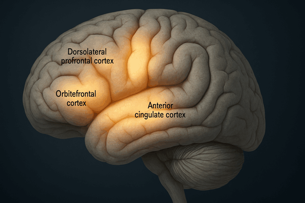
Among the 60 regions of the brain, several reside in the frontal lobe, a region crucial for planning, judgment, problem-solving, and personality expression. The dorsolateral prefrontal cortex, for instance, is heavily involved in working memory and decision-making. It integrates information from sensory and emotional inputs to generate goal-directed behavior. Damage to this region—whether from trauma or neurodegeneration—can manifest as poor impulse control, disorganization, or apathy.
The orbitofrontal cortex, another key area within the frontal lobe, modulates emotional responses and social behavior. It interacts with the amygdala and limbic system to assess the emotional valence of stimuli, influencing everything from empathy to aggression. In a healthy cross-section of human brain anatomy, the connections between these regions demonstrate robust bidirectional communication, facilitating emotional regulation and adaptive behavior. Disruptions here are often implicated in mood disorders, including depression and bipolar disorder.
The anterior cingulate cortex serves as a bridge between emotion and cognition. It detects conflict, monitors performance, and motivates action—a role that makes it essential for attention and focus. Research shows that individuals with underactive anterior cingulate activity often struggle with anxiety and obsessive-compulsive tendencies. Together, the frontal lobe’s contributions illustrate how specific brain regions, when mapped and studied through cross-sectional imaging, illuminate the roots of complex psychological phenomena.
Parietal and Temporal Lobes: Integrating Sensory Experience and Language
Transitioning from the executive command center, the parietal and temporal lobes house numerous regions that mediate sensory integration, language, and memory. In the parietal lobe, the somatosensory cortex receives and processes tactile information, enabling you to perceive touch, pressure, and proprioception. This information is further refined by adjacent association areas that integrate visual and spatial data, which are critical for activities like navigation and hand-eye coordination.
In cases of parietal lobe damage, patients may experience conditions such as hemispatial neglect, where they are unaware of one side of their environment. Understanding the cross-section of human brain structure in this context is crucial for rehabilitation planning and therapeutic interventions. The superior parietal lobule, for instance, is particularly relevant for spatial orientation and attention—skills often impaired in individuals with ADHD or after a stroke.
Meanwhile, the temporal lobe encompasses several of the 60 regions of the brain devoted to auditory processing, memory encoding, and language comprehension. Wernicke’s area, located in the left temporal lobe for most people, is vital for understanding spoken and written language. Lesions here produce receptive aphasia, where speech remains fluent but lacks meaningful content. Adjacent to this is the hippocampus, a seahorse-shaped structure essential for converting short-term memories into long-term storage. Understanding these spatial relationships within cross-section brain anatomy equips clinicians with the knowledge needed to interpret symptoms and devise precise treatment plans.
The Occipital Lobe and Visual Processing Pathways
The occipital lobe, positioned at the back of the brain, is the primary center for visual processing. While it may seem less involved in emotional or cognitive life, its role is foundational to how we interpret the world. Visual information enters through the retina and travels via the optic nerve to the lateral geniculate nucleus, eventually reaching the primary visual cortex located in the occipital lobe. This cortex, also known as V1, is responsible for processing basic visual cues like light intensity, contrast, and orientation.
Beyond this, the visual data is transmitted to secondary and tertiary visual areas that detect motion, depth, and object recognition. These specialized regions within the 60 regions of the brain allow you to identify faces, recognize threats, and navigate environments—all of which feed into emotional and cognitive reactions. A person with damage to the occipital lobe may suffer from cortical blindness or visual agnosia, conditions where sight is preserved but the brain cannot interpret visual stimuli. Understanding how these regions relate within a cross section of human brain anatomy enhances our grasp of how sensory information underpins higher-order thinking.
Limbic System and Emotional Regulation
Diving deeper into the brain’s interior, the limbic system is a cluster of interconnected regions that regulate emotion, motivation, and memory. Within this network, the amygdala acts as the brain’s alarm system, detecting threats and generating fear responses. When hyperactive, it is linked to anxiety and post-traumatic stress disorder. When underactive, emotional blunting or risk-seeking behavior may emerge. Surrounding the amygdala are other essential structures like the hypothalamus and thalamus, which coordinate hormonal responses and sensory routing, respectively.
The hippocampus, also part of the limbic system, not only stores declarative memories but also gives contextual relevance to emotional experiences. It helps you remember where you were when something joyful or traumatic occurred. This interplay between emotion and memory explains why the limbic system is so central to discussions about mental well-being. Disruptions in the limbic circuitry, often visible in cross-section brain anatomy through MRI or PET scans, are frequently observed in individuals with depression, schizophrenia, and neurodegenerative diseases.
Moreover, the limbic system connects extensively with the prefrontal cortex, allowing emotional and cognitive inputs to influence each other. This functional integration—only apparent when examining a cross section of human brain tissue in detail—helps explain why emotional dysregulation often coexists with cognitive symptoms in mood and anxiety disorders.
The Brainstem and Cerebellum: Silent Architects of Stability
Often overshadowed by the cerebral cortex, the brainstem and cerebellum are indispensable for maintaining physiological equilibrium and coordinated movement. The brainstem consists of the midbrain, pons, and medulla oblongata, each of which governs life-sustaining functions like breathing, heart rate, and arousal. Without the brainstem’s efficient relay system, higher brain regions would be cut off from the body’s vital systems.
The cerebellum, nestled beneath the occipital lobe, coordinates voluntary movement, posture, and balance. Once thought to be solely motor-related, emerging research now shows the cerebellum’s involvement in cognitive tasks like attention, language, and even emotional regulation. Within the 60 regions of the brain, specific cerebellar lobules interact with cortical areas to support learning and adaptation.
The cross-section brain anatomy clearly delineates how cerebellar and brainstem structures connect to both spinal and cortical pathways. These insights are particularly useful in cases of neurodegenerative conditions like multiple system atrophy or spinocerebellar ataxia, where damage to these structures leads to both motor and non-motor symptoms. Understanding these foundational regions empowers clinicians and researchers to create holistic treatment strategies that address both the physical and psychological dimensions of health.
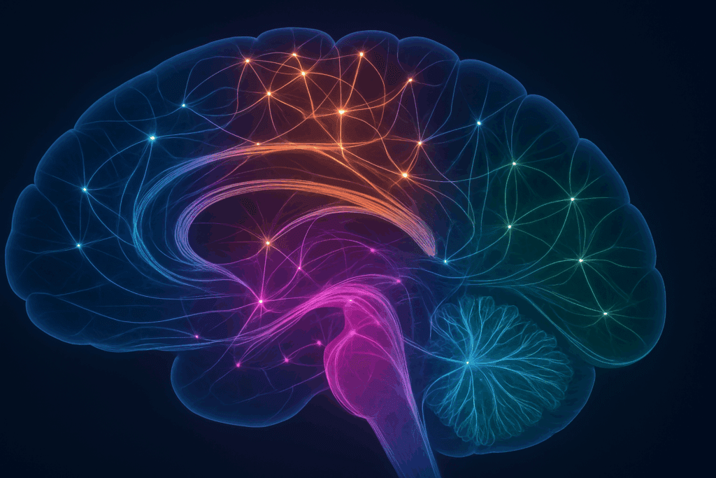
The Connectome: Mapping the Brain’s Functional Network
Beyond discrete anatomical regions, the brain operates as a network—a connectome—composed of billions of synapses and fiber tracts linking the 60 regions of the brain. This connectivity is as critical as the function of individual areas, as it dictates the flow of information and the brain’s adaptive capacity. Functional MRI (fMRI) and diffusion tensor imaging (DTI) are modern tools that visualize this interconnectivity within the cross-section of human brain networks.
The default mode network (DMN), for instance, is active when the brain is at rest and involved in introspection, memory retrieval, and future planning. The salience network, meanwhile, identifies and prioritizes emotionally or cognitively relevant stimuli. Disruptions in these networks—rather than isolated structural damage—are increasingly implicated in conditions like ADHD, autism, and schizophrenia. Understanding cross-section brain anatomy through the lens of network science opens new doors for therapeutic interventions, such as neuromodulation and cognitive rehabilitation.
This networked view does not diminish the importance of the 60 regions of the brain; rather, it enriches it by demonstrating how even small anomalies in connectivity can ripple through broader systems. Whether through trauma, inflammation, or neurodegeneration, disrupted pathways can lead to cascading effects that impair mental clarity, emotional balance, and behavioral regulation.
Frequently Asked Questions: The Cross Section of the Human Brain and Its Impact on Cognitive and Mental Health
Q1. How does understanding the cross section of the human brain influence approaches to mental health therapy?
A detailed understanding of the cross section of human brain anatomy has transformed how mental health therapies are tailored and applied. Rather than relying solely on generalized treatment models, mental health practitioners can now align specific symptoms with functional deficits in the brain’s structure. For instance, targeted cognitive-behavioral therapies for anxiety may focus on enhancing connectivity between the amygdala and the prefrontal cortex, two regions clearly visible and distinct within a brain cross section. As researchers map the 60 regions of the brain more precisely, interventions are becoming more personalized—enabling therapies that match an individual’s neuroanatomical profile. Cross section brain anatomy also informs the development of neurofeedback and transcranial magnetic stimulation (TMS), which rely on pinpointing exact brain regions for modulation, offering a new frontier for non-pharmacological treatment strategies.
Q2. Can lifestyle choices directly influence the way different regions appear in a brain cross section over time?
Yes, consistent lifestyle choices such as diet, physical activity, and cognitive stimulation can produce structural and functional changes observable in a cross section of human brain scans over time. For example, aerobic exercise is known to increase the volume of the hippocampus, a region linked to memory and learning, which appears prominently in cross section brain anatomy. Similarly, mindfulness meditation has been associated with thickening of the anterior cingulate cortex, enhancing emotional regulation. These adaptations highlight neuroplasticity—the brain’s ability to reorganize itself—which is especially relevant when studying the dynamic relationships among the 60 regions of the brain. In clinical imaging, such changes can be tracked longitudinally to assess intervention effectiveness and optimize behavioral health strategies.
Q3. How do the 60 regions of the brain communicate to maintain mental well-being?
The 60 regions of the brain are not isolated entities but are deeply interconnected through functional and structural pathways. This integrative communication is essential for maintaining balanced mood, attention, memory, and emotional resilience. Brain scans using a cross section of human brain imaging reveal how networks like the default mode network (DMN) and the salience network coordinate multiple regions for coherent cognitive performance. These interactions are particularly evident in cross section brain anatomy when evaluating neurological diseases, where even a small disruption in one region can lead to widespread dysfunction. Understanding these collaborative networks also supports the development of pharmacological treatments aimed at modulating network-wide neurotransmitter activity, rather than targeting single areas in isolation.
Q4. What role does cross section brain anatomy play in detecting early cognitive decline or dementia?
Cross section brain anatomy plays a vital role in identifying the subtle signs of cognitive decline before symptoms become clinically significant. Advanced neuroimaging allows clinicians to observe atrophy, reduced blood flow, or changes in metabolic activity within specific regions like the hippocampus and parietal cortex—two of the 60 regions of the brain closely associated with early Alzheimer’s disease. By examining a detailed cross section of human brain scans, practitioners can detect asymmetries or abnormalities that signal neurodegeneration long before cognitive impairment becomes outwardly apparent. This early detection facilitates timely interventions, such as lifestyle changes or cognitive training programs, which can slow progression and preserve mental functioning.
Q5. Are there any practical applications of cross section brain anatomy in educational environments?
Absolutely. Educators, particularly those working with neurodiverse populations, can leverage insights from cross section brain anatomy to tailor teaching strategies that align with the cognitive strengths and weaknesses of their students. For instance, recognizing that the prefrontal cortex plays a critical role in executive function, educators might design classroom activities that support planning, self-monitoring, and emotional control. As research into the 60 regions of the brain deepens, educational psychologists can identify patterns in learning disabilities that correspond to specific anatomical differences, such as underactivity in the parietal lobes linked to dyscalculia. Moreover, brain-based learning programs now use cross section of human brain insights to create more engaging and cognitively compatible learning experiences.
Q6. How is artificial intelligence being used to interpret the cross section of human brain scans?
Artificial intelligence (AI) is rapidly transforming the field of neuroimaging by automating the analysis of complex cross section of human brain data. Machine learning algorithms can identify patterns in large datasets that would be difficult for human observers to detect, including subtle anomalies in the 60 regions of the brain. These tools can classify brain regions, segment tissues, and even predict mental health conditions based on neural architecture captured in cross section brain anatomy. For example, AI-driven diagnostics can assess hippocampal volume to estimate dementia risk or analyze connectivity patterns to screen for depression or autism spectrum disorder. The growing integration of AI enhances both accuracy and speed in clinical decision-making, pushing the boundaries of personalized brain health.
Q7. How do hormonal changes impact the 60 regions of the brain, particularly in women?
Hormonal fluctuations, especially during puberty, menstruation, pregnancy, and menopause, exert significant influence on brain function and structure. These hormonal waves impact several of the 60 regions of the brain, including the hypothalamus, amygdala, and prefrontal cortex, all of which are visible in detailed cross section brain anatomy scans. Estrogen, for instance, has been shown to enhance synaptic connectivity and neuroplasticity in the hippocampus, improving memory during certain phases of the menstrual cycle. Conversely, hormonal imbalances can lead to mood disturbances, brain fog, or anxiety, depending on which brain regions are affected. Understanding the cross section of human brain in the context of endocrinology enables more nuanced treatment approaches for mental health conditions with hormonal underpinnings.
Q8. What are some emerging research areas exploring the 60 regions of the brain?
Current research is increasingly focused on decoding how microstructural differences in the 60 regions of the brain relate to complex behaviors, psychological resilience, and susceptibility to mental illness. One promising area involves mapping individual variability in cross section brain anatomy to predict behavioral traits and cognitive performance. Researchers are also investigating how environmental factors like pollution or chronic stress can induce long-term changes in brain structure, with particular attention to limbic and prefrontal areas. Additionally, psychedelic-assisted therapy is gaining scientific attention, as it appears to temporarily alter connectivity between specific brain regions, an effect that is being quantified through advanced cross section of human brain imaging techniques. These studies are reshaping how we think about neural function, therapy, and the potential for long-term brain health optimization.
Q9. Can abnormalities in cross section brain anatomy explain certain personality traits or tendencies?
There is growing interest in the relationship between structural brain differences and personality traits. Variations in cross section of human brain images often reveal subtle differences in the thickness or volume of areas such as the ventromedial prefrontal cortex or anterior insula—regions known to influence empathy, risk aversion, or impulsivity. While these findings should be interpreted cautiously and not deterministically, there is evidence suggesting that the configuration of the 60 regions of the brain may contribute to consistent behavioral patterns. Longitudinal studies are now examining how life experiences, environment, and genetic predispositions shape cross section brain anatomy over time, providing insights into why people think, feel, and act differently. Such research deepens our understanding of the interplay between biology and personality without reducing identity to mere brain scans.
Q10. How can knowledge of cross section brain anatomy support aging populations in maintaining cognitive health?
As the global population ages, preserving cognitive health has become a major public health priority. Knowledge of cross section brain anatomy allows geriatric specialists to better understand which regions of the brain are most vulnerable to age-related decline and how to protect them. For instance, the hippocampus and prefrontal cortex—both among the 60 regions of the brain—tend to shrink with age, leading to memory lapses and decreased executive function. With insights gained from studying a cross section of human brain imaging, tailored interventions like targeted physical exercise, mental enrichment activities, and dietary strategies can be implemented to support neuroplasticity. This anatomical awareness fosters more proactive, individualized aging plans that prioritize both mental acuity and quality of life.
Conclusion: Why Understanding Cross-Section Brain Anatomy Matters for Mental Health
As we’ve seen, exploring the cross section of human brain anatomy is not merely an academic exercise—it’s a practical pathway to improving mental health outcomes and enhancing cognitive well-being. Each of the 60 regions of the brain contributes to a finely tuned symphony of thought, emotion, memory, and behavior. From the executive powers of the prefrontal cortex to the threat-detection mechanisms of the amygdala and the silent stabilizing role of the cerebellum, these regions interact in deeply integrated ways.
By understanding how these areas are arranged and how they communicate within a cross-section brain anatomy framework, clinicians can better diagnose and treat neurological and psychiatric conditions. Patients, too, can gain insight into how lifestyle choices—such as exercise, sleep, meditation, and nutrition—can influence specific regions or networks within the brain. This anatomically informed perspective encourages a holistic, science-driven approach to brain health that respects both complexity and individuality.
In an era where mental health challenges are increasingly prevalent, the value of understanding the brain’s inner workings has never been higher. Whether you’re interpreting an fMRI scan, engaging in mindfulness to strengthen the anterior cingulate, or adjusting your lifestyle to support hippocampal resilience, knowledge of the brain’s cross-sectional anatomy becomes a guide—not just for understanding disease but for optimizing wellness. Embracing this knowledge allows both professionals and individuals to navigate the intricacies of brain function with clarity, purpose, and hope.
Further Reading:
How does brain geometry influence human brain function?
