Introduction
The human brain is a marvel of biological engineering, orchestrating countless functions that govern our thoughts, movements, and interactions with the world. Among its many vital components, the brain stem and cerebellum play an indispensable role in maintaining balance, coordination, and smooth motor function. These structures form a highly interconnected system that processes sensory input, integrates motor commands, and fine-tunes movement to ensure precision and fluidity. Understanding their functions and interdependence sheds light on the complexities of human motion and helps in diagnosing and treating disorders that affect coordination and equilibrium.
You may also like : Best Things for Brain Health: Expert-Backed Strategies to Keep Your Mind Sharp
Anatomy of the Brain Stem and Cerebellum
The Brain Stem: Structure and Function
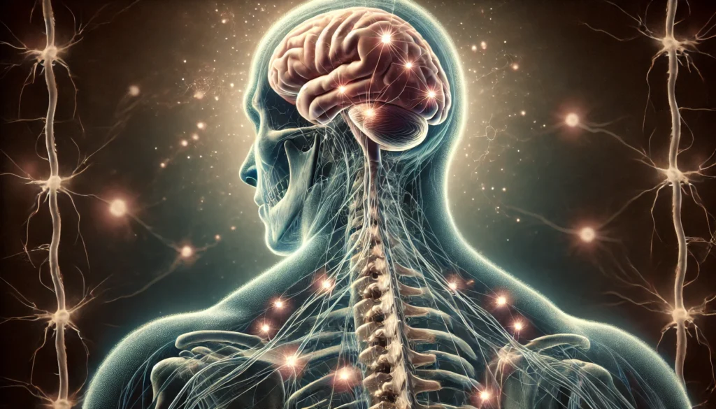
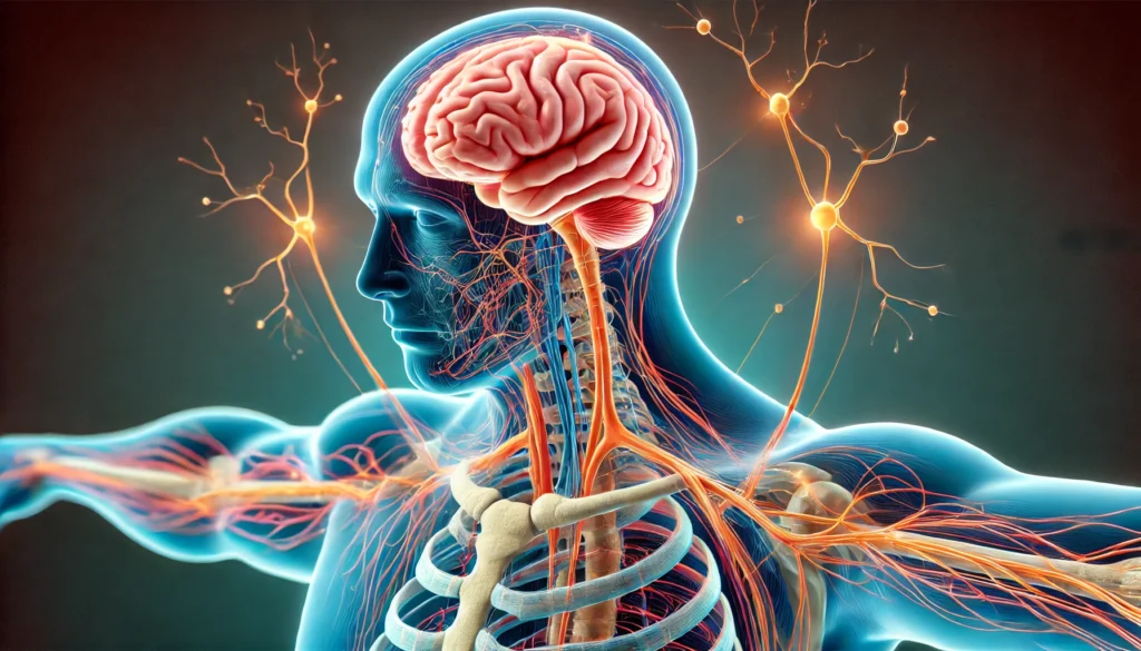
The brain stem is the conduit between the brain and the spinal cord, serving as a communication highway for neural signals. It is composed of three primary sections: the midbrain, pons, and medulla oblongata. These regions are collectively responsible for essential autonomic functions such as respiration, heart rate regulation, and reflexive movements. The brain stem location at the base of the brain makes it a critical structure for survival, acting as the primary gateway for sensory and motor signals traveling between the brain and the rest of the body.
Each part of the brain stem contributes uniquely to motor control. The midbrain is heavily involved in voluntary movement and the processing of visual and auditory stimuli. The pons serves as a major relay center, transmitting information between the cerebrum and cerebellum, while also playing a role in postural stability. The medulla oblongata houses nerve centers responsible for involuntary processes like breathing and swallowing, both of which are necessary for coordinated movement. Understanding these brain stem parts and functions provides critical insights into how the body maintains movement precision and balance.
The Cerebellum: Structure and Function
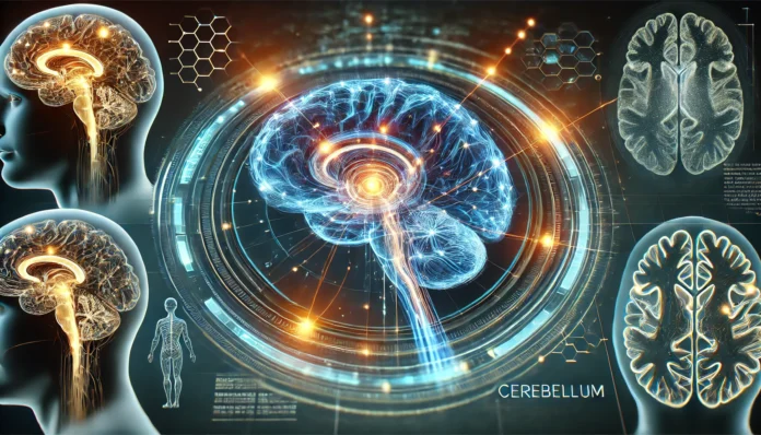
The cerebellum, often referred to as the “little brain,” is located at the back of the skull, just beneath the occipital lobe. When examining brain anatomy, the cerebellum stands out as a highly folded structure that contains nearly half of the brain’s total neurons. The cerebellum of the brain is primarily responsible for fine-tuning motor activity, ensuring movements are smooth, coordinated, and accurate.
The cerebellum is divided into several distinct regions known as lobes. The anterior and posterior lobes work together to refine voluntary movements, while the flocculonodular lobe is instrumental in maintaining balance and spatial orientation. By continuously processing input from the inner ear, eyes, and proprioceptors in muscles and joints, the cerebellum helps the body adjust movements in real-time. This function of the cerebellum is crucial in everyday activities such as walking, running, and grasping objects.
The Interplay Between the Brain Stem and Cerebellum
Neural Communication Pathways
The brain stem and cerebellum function as a unified system, linked by intricate neural circuits that allow for the seamless execution of motor tasks. Neural signals travel through the pons, which serves as the primary connection between the cerebellum and the cerebral cortex. The information exchanged through these pathways ensures that voluntary movements initiated in the motor cortex are refined for precision and efficiency.
One of the most critical aspects of this neural interplay is the cerebellum’s ability to detect and correct errors in movement. If an individual missteps or loses balance, the cerebellum processes this information instantaneously and sends corrective signals to the brain stem, which then adjusts muscle activity accordingly. This rapid feedback loop is fundamental in preventing falls and maintaining equilibrium.
Coordination and Motor Learning
The integration of brain stem and cerebellum function extends beyond simple movement correction—it also plays a vital role in motor learning. The cerebellum is essential in refining learned motor skills through repeated practice. For example, when learning to ride a bicycle, the cerebellum and brain stem work together to stabilize posture, adjust muscle coordination, and adapt movements based on sensory feedback.
Studies on cerebellum brain function indicate that damage to these structures can lead to motor impairments such as ataxia, where coordinated movement becomes difficult or impossible. The brain stem cerebellum connection is therefore critical for activities that require dexterity and fluidity, from typing on a keyboard to playing a musical instrument.

The Role of the Brain Stem and Cerebellum in Balance and Coordination
Maintaining Postural Stability
One of the most fundamental questions in neurobiology is: what part of the brain controls balance and coordination? The answer lies within the cerebellum and brain stem. These structures process vestibular input from the inner ear and proprioceptive signals from muscles and joints to maintain postural control. The cerebellum ensures that equilibrium is preserved by continuously adjusting muscle tone and limb positioning in response to external stimuli.
For instance, when an individual stands on an uneven surface, the brain stem cerebellum network rapidly analyzes sensory data and adjusts muscle contractions to stabilize the body. This complex interaction prevents falls and allows humans to navigate their environment with confidence.
Reflexive and Voluntary Movements
Beyond basic postural adjustments, the brain stem is also involved in reflexive and voluntary motor functions. Reflexive actions, such as quickly pulling a hand away from a hot surface, are mediated by the brain stem’s automatic response systems. In contrast, voluntary movements, such as reaching for a cup of coffee, involve a dynamic collaboration between the cerebellum and the motor cortex, facilitated through brain stem pathways.
Clinical Implications and Disorders
Neurological Disorders Affecting Balance and Coordination
Damage to the brain stem or cerebellum can result in a range of movement disorders, significantly affecting an individual’s ability to perform daily tasks. Conditions such as cerebellar ataxia, Parkinson’s disease, and multiple sclerosis often involve impairments in balance and coordination due to disruptions in these neural circuits.
Cerebellar ataxia, for example, manifests as difficulty with coordinated movements, resulting in an unsteady gait and problems with fine motor tasks. Similarly, lesions in the brain stem can affect cranial nerve function, leading to issues with eye movement control and overall motor precision.
Rehabilitation and Treatment Strategies
Advances in neurorehabilitation have focused on targeting the brain stem cerebellum system to restore lost function. Physical therapy interventions, such as balance training and coordination exercises, help reinforce neural pathways and improve motor control. Research into neuroplasticity suggests that targeted rehabilitation can enhance recovery in patients with cerebellar dysfunction by strengthening alternative neural connections.

Frequently Asked Questions (FAQ): How the Brain Stem and Cerebellum Work Together
1. How does the brain stem cerebellum connection impact everyday movement? The brain stem cerebellum connection is fundamental for executing smooth and coordinated movements in daily life. Whether walking, writing, or even standing still, this system ensures that muscles work in harmony to prevent awkward or jerky motions. Without a properly functioning brain stem cerebellum network, movements would lack fluidity, making tasks like reaching for a glass or stepping off a curb difficult. The cerebellum of the brain refines motion by integrating sensory feedback, while the brain stem acts as the relay center for motor commands. This coordination allows the body to adapt to changing environments and maintain precision in movement.
2. Where is the cerebellum located, and why is its position significant? The cerebellum location at the base of the skull, just above the brain stem, is crucial for its function. It is positioned strategically to receive information from both the spinal cord and the higher brain regions, allowing it to process movement-related data efficiently. By being closely connected to the brain stem, it can quickly send corrective motor signals to adjust posture and coordination. Understanding where the cerebellum is located in the brain helps researchers and medical professionals diagnose and treat disorders affecting motor function. This placement also makes the cerebellum vulnerable to injuries from trauma or degenerative diseases that impact balance and coordination.
3. What parts of the brain stem are most involved in motor coordination? Motor coordination primarily involves the three parts of the brain stem: the midbrain, pons, and medulla oblongata. The midbrain processes sensory input and initiates motor responses, playing a key role in reflexive movements. The pons acts as a bridge between the cerebellum and the cerebrum, ensuring that voluntary movements are refined and accurate. The medulla oblongata is responsible for controlling involuntary movements such as breathing and heartbeat, which indirectly affect balance and muscle tone. These three regions work in unison to ensure that the function of the brain stem in brain activity remains efficient and precise.
4. How does the brain stem diagram illustrate its function in movement?
A brain stem diagram typically shows the three primary sections of the brain stem and their roles in movement and coordination. By visualizing the parts of the brain stem, it becomes clear how different regions contribute to motor control, balance, and reflexes. The labeled brain stem in such diagrams often highlights how sensory and motor pathways travel through the structure, reinforcing its role as a communication hub. Understanding brain anatomy brain stem relationships through these diagrams is essential for medical education and research. These visual tools help pinpoint where dysfunctions occur in conditions like stroke, Parkinson’s disease, and other movement disorders.
5. What part of the brain controls balance and coordination, and how does it function?
The cerebellum is the primary structure responsible for balance and coordination, working closely with the brain stem. It processes sensory information from the inner ear, muscles, and eyes to make real-time adjustments to posture and movement. The brain stem cerebellum connection is vital for executing coordinated movements, ensuring that balance is maintained even in dynamic activities like running or dancing. Without a functioning cerebellum, individuals may experience unsteady movements, poor muscle control, and difficulty adapting to changes in terrain. By continuously fine-tuning motor commands, the cerebellum enables smooth, purposeful motion.
6. How many regions make up the brain stem, and how do they interact with the cerebellum?
The brain stem consists of three primary regions: the midbrain, pons, and medulla oblongata. Each of these regions interacts with the cerebellum to regulate movement and posture. The pons, in particular, serves as a major communication bridge, transmitting signals between the cerebellum of the brain and higher cortical areas. The midbrain facilitates quick reflexive movements based on visual and auditory cues, while the medulla oblongata ensures involuntary movements remain stable. Understanding what makes up the brain stem helps in comprehending its intricate relationship with the cerebellum and its role in controlling muscle coordination.
7. What happens if the cerebellum or brain stem is damaged?
Damage to the brain stem or cerebellum can result in severe impairments in balance, coordination, and motor function. Depending on which parts of the brain stem are affected, symptoms can range from loss of fine motor control to complete loss of movement in certain areas of the body. Conditions like stroke, traumatic brain injuries, or neurodegenerative diseases can compromise the function of brain stem in brain activities, leading to difficulties in walking, speaking, or even maintaining posture. In severe cases, damage to these regions can affect involuntary functions like breathing and heart rate, making early diagnosis and intervention crucial. Rehabilitation therapies often focus on retraining the brain to compensate for lost functions, utilizing neuroplasticity to enhance recovery.
8. How does the cerebellum brain function support motor learning?
The cerebellum is not just involved in coordinating movement; it also plays a key role in motor learning. When learning a new physical skill, such as playing an instrument or riding a bicycle, the cerebellum stores motor patterns that allow movements to become more refined with practice. The brain stem cerebellum connection ensures that adjustments are made in real-time, helping to correct errors and improve efficiency. This process is crucial in sports, where muscle memory and fine motor control are required for peak performance. The cerebellum lobe structures contribute to refining and adapting motor commands to enhance overall movement efficiency.
9. What parts of the brain make up the brain stem, and how do they regulate movement?
When considering what parts of the brain make up the brain stem, the midbrain, pons, and medulla oblongata stand out as essential regions. These structures regulate movement by transmitting signals between the brain and the body, ensuring voluntary and involuntary functions are executed properly. The brain diagram brain stem visualizes these interactions, showing how nerve pathways integrate motor and sensory information. A labeled brain stem can highlight specific areas where movement control is initiated and refined. The brainstem diagram further explains how neural circuits facilitate coordination with the cerebellum, ensuring fluid motion and posture control.
10. Why is the cerebellum often referred to as the “little brain,” and what does it control?
The cerebellum is often called the “little brain” because it has a highly folded structure similar to the cerebrum but is much smaller in size. Despite its compact form, it plays a critical role in coordinating voluntary movements, fine-tuning motor control, and maintaining balance. The cerebellum location within the posterior cranial fossa positions it perfectly to receive input from the spinal cord, sensory organs, and cerebral cortex. Understanding what the cerebellum is responsible for helps medical professionals diagnose conditions that affect movement precision and balance. By continuously adjusting motor output, the cerebellum ensures that actions are carried out smoothly and with precision.

Conclusion
The synergy between the brain stem and cerebellum is essential for balance, coordination, and motor precision. Their intricate neural pathways facilitate smooth movement execution and rapid adjustments in response to sensory input. Understanding the cerebellum location and its connection to brain stem structures provides insight into the foundational mechanisms of motor control. As research advances, exploring therapeutic approaches for disorders affecting these regions will be crucial in improving mobility and quality of life for individuals with neurological impairments. The brain anatomy brain stem and cerebellum network remains a cornerstone of movement science, underscoring the importance of this remarkable biological system.
neuroscience of movement, motor control in the brain, balance and posture regulation, coordination disorders, cerebellar function, brain structure and motor skills, neurological pathways, brain regions and motor function, equilibrium and motion, neuroplasticity and movement, central nervous system control, fine motor coordination, motor learning and adaptation, sensory integration in movement, involuntary motor control, cerebellar ataxia insights, proprioception and balance, brainstem connectivity, movement disorders research, neural circuits in motor function
Further Reading:
Which Parts of the Brain Are Responsible for Balance and Equilibrium?
Cerebellum: Anatomy and Function
Disclaimer
The information contained in this article is provided for general informational purposes only and is not intended to serve as medical, legal, or professional advice. While Health11News strives to present accurate, up-to-date, and reliable content, no warranty or guarantee, expressed or implied, is made regarding the completeness, accuracy, or adequacy of the information provided. Readers are strongly advised to seek the guidance of a qualified healthcare provider or other relevant professionals before acting on any information contained in this article. Health11News, its authors, editors, and contributors expressly disclaim any liability for any damages, losses, or consequences arising directly or indirectly from the use, interpretation, or reliance on any information presented herein. The views and opinions expressed in this article are those of the author(s) and do not necessarily reflect the official policies or positions of Health11News.


